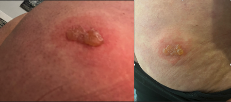
Case Presentation: A 42-year-old female with a past medical history of hereditary angioedema, hypertension, and asthma presented to the emergency department with sudden onset weakness and numbness in the right upper and lower extremities, coupled with a posterior “pressure-like” headache. On presentation, she was non-febrile, hypertensive at 169/92 mmHg with a regular heart rate (80 beats per minute) and preserved oxygen saturation on room air. Physical examination revealed 3 out of 5 weakness of the right upper and lower extremities, intact sensory function, brisk reflexes, and a negative Babinski sign. A painful right buttocks lesion was noted. (Figure 1) A stroke code was initiated due to the acute neurological symptoms, and aspirin loading was administered. A computed tomography (CT) scan showed no intracranial abnormalities, and a CT angiogram ruled out large vessel occlusion or arterial stenosis. Subsequent MRI revealed subtle scattered subcortical white matter changes over the bilateral frontal and parietal lobes. Upon further historical review, the patient had a history of two shingles flares with right buttock involvement and intermittent neck stiffness. Considering the clinical presentation and negative initial stroke work-up, a presumptive working diagnosis of meningitis was made. Consultation with Infectious Disease led to the recommendation of high dose valacyclovir for ten days based on a high suspicion of Mollaret’s meningitis. HSV 1 and 2 PCR swabs from the buttocks lesion returned positive. The patient improved and was discharged with life-long prophylactic anti-viral therapy and home physical therapy. All symptoms resolved by the two-week follow-up appointment.
Discussion: Mollaret’s meningitis is an uncommon condition marked by recurrent aseptic lymphocytic meningitis episodes followed by symptom-free intervals. (1) Symptoms include neck stiffness, headache, nausea, vomiting, photophobia, myalgias, and fevers. Neurological manifestations may encompass visual disturbances, speech impairment, facial paresis, reflex abnormalities, hallucinations, and even coma. Importantly, symptoms typically resolve spontaneously within weeks without residual effects. (2) Our patient presented with acute neurological deficits, intermittent neck stiffness, and a recurrent rash, all historically resolving within weeks without residual neurological sequelae. Diagnosing Mollaret’s syndrome remains a challenge due to the lack of specific tests, relying on clinical symptoms, laboratory tests, and imaging studies for exclusion. (3) Treatment primarily involves supportive measures, but some literature suggests potential benefits from long-term prophylaxis in Mollaret’s meningitis patients. (4)
Conclusions: Our case highlights the rarity and diagnostic challenges of Mollaret’s meningitis, especially when presenting with symptoms resembling a stroke. Increased awareness of this condition is crucial for accurate diagnosis. Ongoing monitoring and management are essential in navigating the complexities of such cases, emphasizing the need for collaboration among healthcare professionals to optimize patient outcomes.