Case Presentation: A 61 year old African American man with past medical history of hyptertension, diabetes mellitus type 2, chronic kidney disease stage 3, remote intravenous drug use, Hepatitis C presented with worsening hypertension, new onset bilateral lower extremity swelling and worsening kidney function since one month. Patient reported weight gain and urinary frequency at home but denied flank pain, hematuria, abdominal pain or fevers. Physical exam was significant for hypertension and 1+ pitting, nonerythematous, nontender edema to the knees bilaterally. Laboratory data on this admission revealed normocytic anemia, creatinine of 2.0 mg/dL (baseline around 1.3 mg/dL), albumin 2.0 g/dL (baseline 4.0 g/dL). Urinalysis showed total protein of 1051 mg/dL, but no hematuria. Serum and urine protein electrophoresis were positive for monoclonal spike (IgG lambda). Serum free light chain assay showed elevated kappa and lambda light chains (lambda more than kappa). Serologic work up was negative as were cryoglobulins. A kidney biopsy performed showed membranoproliferative glomerulonephritis with focal global glomerulosclerosis as well as moderate tubular atrophy and interstitial fibrosis. Immunoflorescence revealed IgM, kappa and lambda staining in a global peripheral fine granular pattern. Bone marrow aspirate and biopsy showed 1% lambda light chain-restricted plasma cells most consistent with MGUS. Nephrology and Hematology/Oncology services were consulted. The presence of myeloma like picture on kidney pathology despite lack of full blown multiple myeloma prompted the decision to initiate therapy targeted towards underlying plasma cell dyscrasia. Patient showed good clinical (resolution of edema and normalization of creatinine) and histopathological response with Cyclophosphamide, Bortezemib and Dexamethasone.
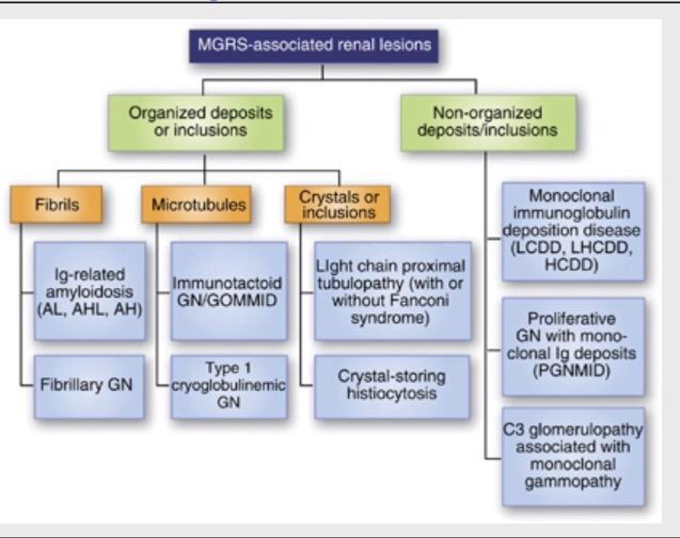
Discussion: Monoclonal Gammopathy of Undetermined Significance (MGUS) is an asymptomatic premalignant clonal plasma cell or lymphoplasmacytic proliferative disorder and occurs in over 3 percent of the general population over the age of 50 and is typically detected as an incidental finding. It is also characterized by lack of lytic bone lesions, anemia, hypercalcemia, and renal insufficiency related to the plasma cell proliferative process. We report a case of membranoproliferative glomerulonephritis caused by monoclonal gammopathy. This condition is now categorized as Monoclonal Gammopathy of Renal Significance (MGRS). The prevalence of clinical MGRS is up to 23% among patients with MGUS.
Conclusions: MGRS is diagnosed by demonstration of monoclonal deposits in the kidney. Immunoflorescence study should be performed on all suspected cases. Restriction to a single class of light chain and/or heavy chain is mandatory. Early recognition is crucial, as suppression of monoclonal immunoglobulin secretion by chemotherapy often improves outcomes. Recovery of renal function is possible with adequate hematologic response. The term MGUS should be limited to those cases where no end organ damage can be demonstrated. Meanwhile, MGRS should be used when the monoclonal protein is playing a direct role in the kidney disease. These disorders do not require treatment from a “tumoral” viewpoint, but treatment is often mandatory and sometimes urgent to prevent renal deterioration.