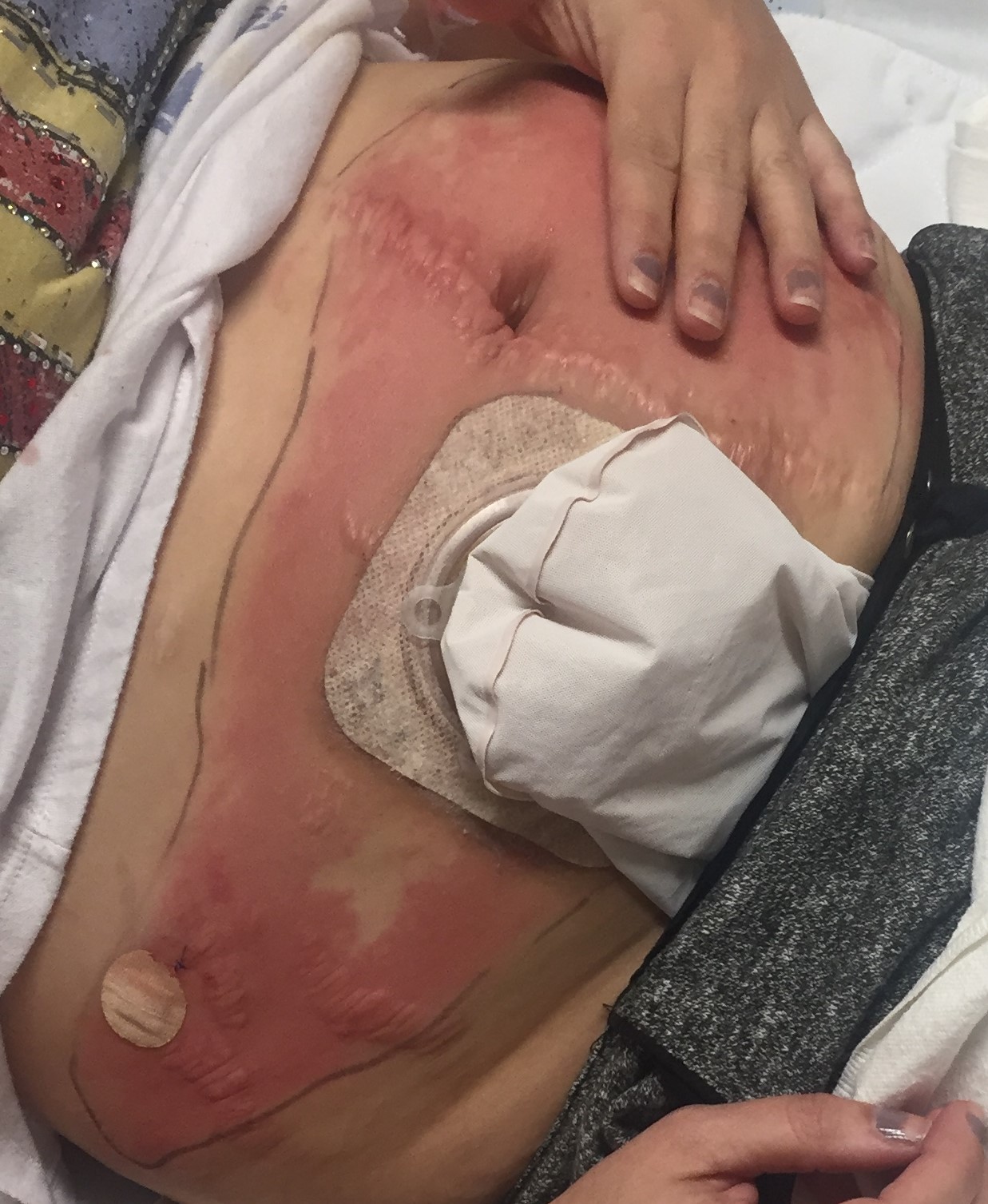
Case Presentation: A 36 year old female, with known ileocolonic Crohn disease, status post total colectomy with end ileostomy, maintained on Infliximab and Azathioprine, presented with one day history of subjective fevers and fixed, non-progressive, abdominal wall rash. Two days prior to presentation she was started on Topiramate and Celecoxib for headaches and joint pain associated with Infliximab infusion, respectively. She denied any new topical creams, lotions, medications, or soaps to the area. She denied any abdominal pain or increased ostomy output. Examination revealed a temperature of 36.7 and a tender, large, well circumscribed, pink, edematous plaque on the lower abdomen, with an ostomy on the right lower abdomen with a pink healthy stoma. On admission, she had leukocytosis to 11.9 with 90% neutrophils and no eosinophilia. Gram stains and blood cultures were negative. CT scan of the abdomen was unrevealing. Infliximab and azathioprine metabolites levels were checked and found to be within therapeutic range. She was started on Clindamycin due to concern for cellulitis with no improvement. Dermatology were consulted and a presumptive diagnosis of fixed drug eruption was made in the setting of new medication use, however, subsequent punch biopsy showed perivascular mixed inflammatory infiltrate with focal granuloma, consistent with metastatic Crohn disease. She was started on Clobetasol 0.05% ointment with significant improvement of her rash. She was discharged on Clobetasol twice daily and a 4 week prednisone taper with complete resolution of her rash on outpatient follow-up. Since her initial presentation, patient had several subsequent admissions for CD flare and recurrence of her MCD, and was considered a primary non-responder to anti-TNF therapy and switched to Ustekinumab with control and sustained remission of her gastrointestinal and cutaneous symptoms.
Discussion: It has been reported that up to 44% of patients with Crohn diseases (CD) experience extraintesitnal manifestations in the form of dermatologic pathologies. Metastatic Crohns disease (MCD) is a rare dermatologic entity with fewer than 100 cases reported in the literature. Its clinical polymorphism and similarity to other disease processes, make its diagnosis challenging. An integration of clinical information, histopathologic findings of noncaseating granulomas at sites anatomically separate from the gastrointestinal tract, and ancillary studies to exclude other diagnosis are necessary for accurate diagnosis. Treatment of MCD remains anecdotal due to the paucity of cases, however success has been reported with various regimens, including antibiotics, immunosuppressants, and sometimes surgery. In our case, sustained remission was achieved with Ustekinumab after failure of combination therapy with steroids and infliximab.
Conclusions: Due to its rarity and wide clinical variability MCD poses a diagnostic and therapeutic challenge. A biopsy should be considered in patients with CD presenting with an unspecified dermatologic lesion to avoid delay in diagnosis and decrease rate of inappropriate therapies.