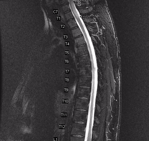
Case Presentation: A 52 year-old man with metastatic esophageal adenocarcinoma undergoing chemoradiation presented with two weeks of asymmetric numbness. On initial presentation, he noted progressive, ascending bilateral lower extremity numbness to the waist. Examination was normal, except for decreased pinprick below T10, loss of vibratory sense below mid-thigh, decreased proprioception of the left toe, positive Romberg sign, and a wide based gait. Cerebrospinal fluid (CSF) analysis demonstrated clear, colorless fluid with an opening pressure of 9 cm H20, lymphocytic predominance with increased histiocytes, no RBCs, normal glucose and protein. MRI of the spine revealed increased T2 signal with ill-defined enhancement within the spinal cord from C6 to T2 without evidence of cord compression, epidural, or leptomeningeal disease. CSF was sent for cytopathology, infectious, and paraneoplastic analysis. He was discharged with follow-up, but returned a few days later with a progressive sensory level to T4 and a papular rash on his medial right thigh, which had not been noted previously. He continued to deny fever, confusion, headache, weakness, back pain, saddle anesthesia, urinary retention, bowel/bladder incontinence, recent travel, vaccinations, sick contacts, or new sexual partners. Cytopathology showed atypical cells in a background of lymphocytes and histocytes, initially interpreted as metastatic disease. However, CSF HSV-2 PCR and serum HSV IgG antibody was noted to be positive. Confirmatory immunostains of the CSF favored an infectious etiology related to HSV-2 rather than a neoplastic process. Additional history revealed that the patient had a genital ulcer three weeks prior that resolved without intervention. He was initiated on acyclovir with significant clinical and radiological improvement.
Discussion: While HSV-2 infections involving the central nervous system typically result in aseptic meningitis, HSV-2 myelitis is a rare cause of myelopathy more prevalent in the immunosuppressed population. It presents as an ascending sensory, motor, or autonomic myelopathy that can progress to necrotizing disease associated with high morbidity and mortality. Distinguishing between malignancy versus HSV-2 related myelopathy can be challenging. In this case, the presence of atypical lymphocytosis and histiocytes within the CSF in addition to a high nuclear to cytoplasm ratio was initially suspected to be related to a malignant process. This demonstrates the diagnostic pitfall of infection mimicking malignancy and reiterates the importance of correlating all clinical data to allow for accurate diagnosis and early intervention. Confirmatory immunostains revealed an infectious etiology rather than malignant, thus leading to appropriate therapy.
Conclusions: Herpes simplex virus (HSV)-2 is a rare cause of myelitis in immunocompromised patients. Given its variable course, rapid diagnosis is essential to prevent permanent disability and death. Cytopathologic features of HSV-2 infection can mimic malignancy, emphasizing the importance of correlating all clinical data.