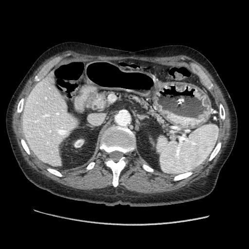
Case Presentation: A 65-year-old male presented to the emergency department with a 5-week history of epigastric pain, nausea, constipation and weight loss. Past medical history was significant for combined liver and kidney transplant 3 years prior due to hepatitis C cirrhosis and hepatorenal syndrome. Chronic immunosuppression consisted of mycophenolate mofetil, tacrolimus and prednisone. Laboratory studies including liver and pancreatic enzymes were unremarkable. Upper GI endoscopy demonstrated significant edema in the duodenal region with partial obstruction of the lumen. CT abdomen was consistent with groove pancreatitis and did not show a pancreatic mass or dilated bile ducts. Repeat endoscopy showed completed obstruction in the first part of the duodenum. An unsuccessful balloon dilation was attempted. Endoscopic ultrasound (EUS) was limited by the obstruction and did not visualize the pancreatic head. Multiple biopsies of the region from both procedures were negative for malignancy. The patient was discharged after placement of a gastrostomy-jejunostomy tube for symptomatic relief and feeding. He was readmitted 2 months later with hematemesis. Endoscopy showed hemorrhagic gastritis with persistent duodenal obstruction requiring surgical gastrojejunostomy. Postoperatively he developed jaundice and abnormal liver function tests. MRI demonstrated a biliary stricture. Endoscopic retrograde cholangiopancreatography (ERCP) demonstrated severe stricture of intrapancreatic bile ducts. Brushing cytology revealed adenocarcinoma. EUS showed a pancreatic head mass and biopsy confirmed a pancreatic adenocarcinoma. Tumor resectability was considered borderline and neoadjuvant chemotherapy was initiated. Unfortunately, liver metastases were detected after 3 cycles and the patient enrolled in hospice.
Discussion: Groove pancreatitis is a rare form of chronic pancreatitis that usually occurs in males in the fourth and fifth decades of life with a history of alcohol abuse. The most common presentation is duodenal obstruction. Most patients have elevated pancreatic enzymes and some develop jaundice. Surgery in the duodenal and biliary duct region may predispose patients to develop the condition, which can be exquisitely hard to distinguish from pancreatic adenocarcinoma, as this case illustrates. Ultimate diagnosis commonly entails a Whipple procedure. The absence of prior alcohol abuse or elevated pancreatic enzymes and an increased risk of malignancy due to immunosuppression were hints to the presence of cancer in this patient. His history of extensive abdominal surgery and chronic immunosuppression made him a poor candidate for surgical intervention, further delaying the diagnosis.
Conclusions: This case demonstrates that differentiating between groove pancreatitis and cancer may be extremely difficult. Clinicians should have a high index of suspicion for pancreatic adenocarcinoma in patients presenting with clinical features of groove pancreatitis, especially considering the rarity of the latter diagnosis.