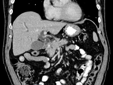
Case Presentation: A 65-year-old businessman with no significant past medical history presented with 2 weeks of epigastric and right upper quadrant (RUQ) abdominal pain. He also noticed the darkening of the urine and pale stools. He had scleral icterus, and abdominal examination revealed no tenderness to palpation. Laboratory tests were notable for total bilirubin 14.8 mg/dL (direct 11.9 mg/dl), AST 189 U/L, ALT 458 U/L, alkaline phosphatase 397 U/L. A RUQ ultrasound showed biliary dilation. A CT abdomen revealed a large pancreatic head mass favored to represent pancreatic ductal adenocarcinoma (figure). He underwent ERCP for a pancreatic mass needle biopsy. A stent was placed for a common bile duct stricture with upstream dilation. An IgG4 level was subsequently ordered and was slightly elevated to 135.9 mg/dL (normal 6-130 mg/dL). The CA19-9 tumor marker was elevated to 670 U/mL (normal 0-40 mg/mL). He was discharged with close oncology and surgical oncology follow-up for treatment of presumed pancreatic cancer.He was readmitted 2 days later for acute cholangitis that required replacement of his occluded biliary stent and treatment with antibiotics. The pathology on the pancreatic mass biopsy was negative for adenocarcinoma and consistent with autoimmune pancreatitis with a notable increase in the number of IgG4 cells. The patient was started on oral prednisone 40 mg daily for 4 weeks for IgG4 related disease (IgG4-RD). A follow-up MRI abdomen showed no active pancreatic inflammation, and resolution of previous mass and biliary dilation
Discussion: Autoimmune pancreatitis (AIP) secondary to IgG4-RD should be included in the differential diagnosis of pancreatic cancer. The biliary stricture seen on ERCP, in this case, prompted suspicion because the combination of biliary tract and pancreatic disease is nearly diagnostic of IgG4-RD. Patients commonly present with painless jaundice and pancreatic tumors. Because of similarities in clinical and radiographic findings, it is often difficult to differentiate between AIP and pancreatic adenocarcinoma, and the treatment of the two etiologies is starkly different. AIP usually responds to steroids, whereas treatment for pancreatic cancer involves surgery and chemotherapy. Therefore, it is vital to establish a diagnosis before initiating therapy. CA19-9 is a tumor marker for pancreatic cancer. There have been several studies investigating CA19-9 cutoff levels. One study reported that in patients with mass lesions, a CA19-9 level greater than 300 U/mL was indicative of malignancy in all cases. Another study showed an elevated CA19-9 to have an overall sensitivity of 81% and specificity of 90% for pancreatic cancer. Serum IgG4 is another biomarker that has been used to help diagnose AIP. A two-fold increase of IgG4 (using a 140 mg/dL cutoff) showed a sensitivity of 53% and specificity of 99% for AIP. However, a mild increase in IgG4 can be seen in up to 10% of patients with pancreatic malignancy.
Conclusions: When evaluating pancreatic head masses, an elevated CA 19-9 does not confirm pancreatic cancer even in the presence of supportive radiographic findings. Given vastly different treatment options, it is essential to also consider AIP secondary to IgG4-RD in the differential diagnosis of a pancreatic mass and the diagnosis confirmed with a biopsy.