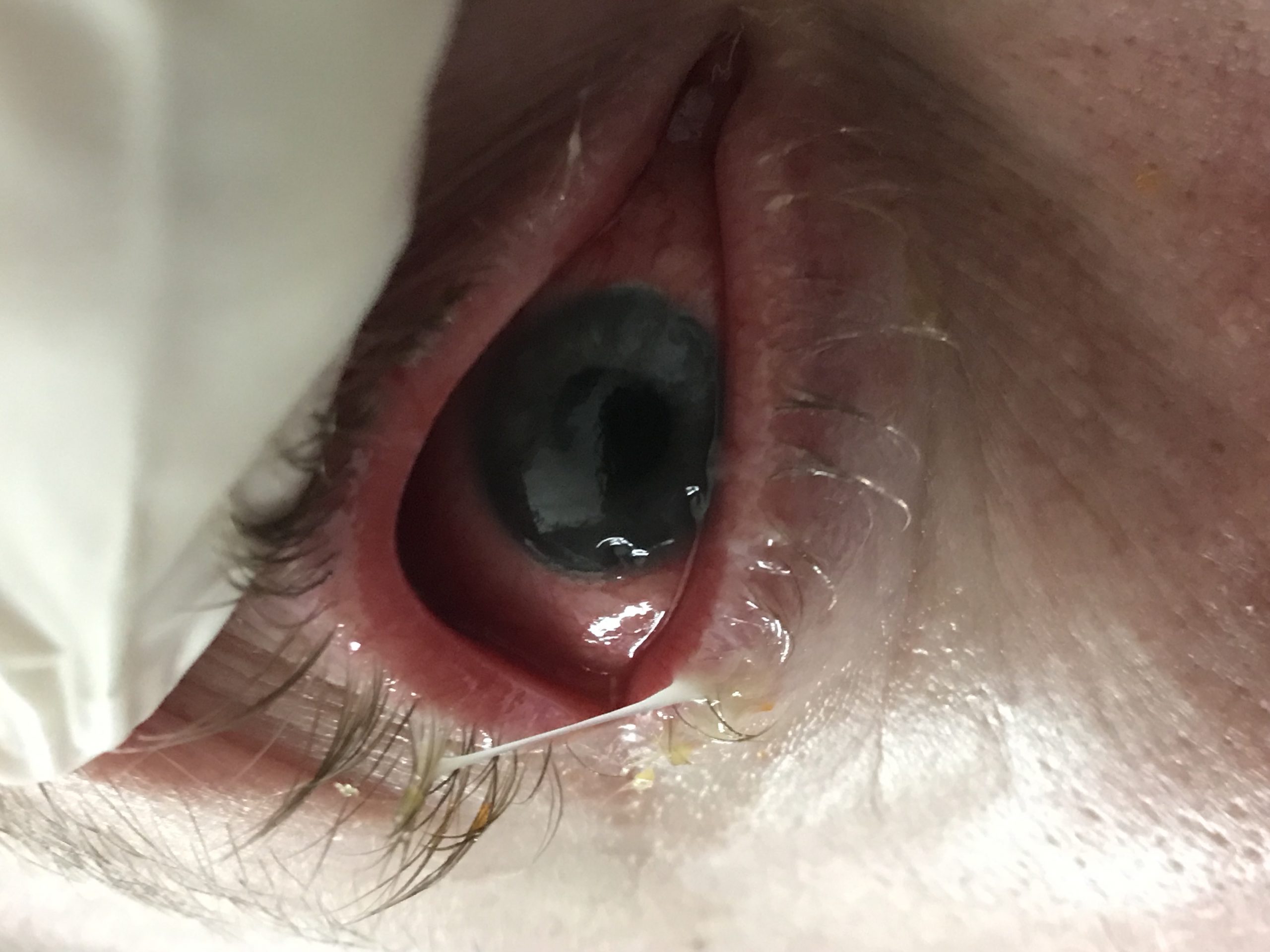
Case Presentation: A 34 year old Caucasian male with a past medical history of untreated Crohn’s disease for 7 years, treatment naïve Hepatitis C, and intravenous drug abuse presented with a month history of gradually worsening bilateral eye pain and drainage. Physical exam revealed severe injection with drainage from conjunctiva and sclera of both eyes. Corneal examination revealed circular areas of ulceration in the inferotemporal and superonasal regions near limbus with mild thinning and vascularization in the right eye; inferonasal crescent with mild thinning and superotemporal round ulceration with severe thinning and descemetocele was observed in the left eye. Due to initial concern for bilateral gonococcal and chlamydial conjunctivitis, the patient was administered a dose of 1 gm oral azithromycin with daily 1 gm intravenous ceftriaxone. Oral doxycycline 100 mg twice daily was added for prevention of further collagen damage; moxifloxacin and tobramycin ophthalmic drops were also provided. After patient failed to improve within 48 hours, vancomycin was included for methicillin resistant Staphylococcus aureus coverage and ceftriaxone was escalated to cefepime for Pseudomonas coverage. Eye and blood cultures remained negative. Rheumatologic tests (anti-Sm, anti-RNP, anti-SSA, anti-SSB, C-ANCA, p-ANCA, RF, and CCP) were negative; other tests including HIV, syphilis, CMV, EBV, and Quantiferon Gold also resulted negative. Curiously, colonoscopy precluded Crohn’s disease flare. Due to lack of improvement with broad spectrum antibiotics for 7 days, daily 1 gm intravenous methylprednisolone for 3 days was administered, which yielded significant improvement in symptoms. Oral prednisone 80mg daily, to be tapered over 2 weeks, as well as, subcutaneous adalimumab 80 mg every 2 weeks were initiated. At the time of discharge patient had significant improvement in eye pain with evidence of corneal re-epithelialization.
Discussion: Inflammatory bowel disease (IBD) is associated with several ophthalmologic sequelae including episcleritis, scleritis and uveitis. Peripheral ulcerative keratitis (PUK) is an extremely uncommon disease with overall incidence of 3 persons per million per year. Moreover, PUK in Crohn’s disease has rarely been described. PUK involves crescent shaped stromal inflammation of juxtalimbal cornea characterized by sectoral thinning of the affected area. Potential etiologies of PUK are infections (Staphylococcus, Streptococcus, and Gonococcus, among others), autoimmune and vasculitides (lupus, and rheumatoid arthritis, among others), and ocular trauma. Lack of treatment may cause progression to corneal perforation and blindness. Initial appearance may resemble conjunctivitis, which may resolve with much simpler management strategies. However, any delay in management of PUK can be sight threatening. Initial treatment comprises of systemic steroids; lack of response may be an indication for immunosuppressant use. Moreover, a few studies have suggested use of biologics. Likewise, management approach in our patient was similar as initial treatment IV solumedrol was followed by oral corticosteroids, and ultimately by adalimumab.
Conclusions: PUK is potentially a sight threatening disease that needs early diagnosis and treatment. Ophthalmology evaluation is recommended for all patients with inflammatory bowel disease, especially prior to medication changes. Usual treatment of PUK involves steroids, immunosuppressants and biologics.