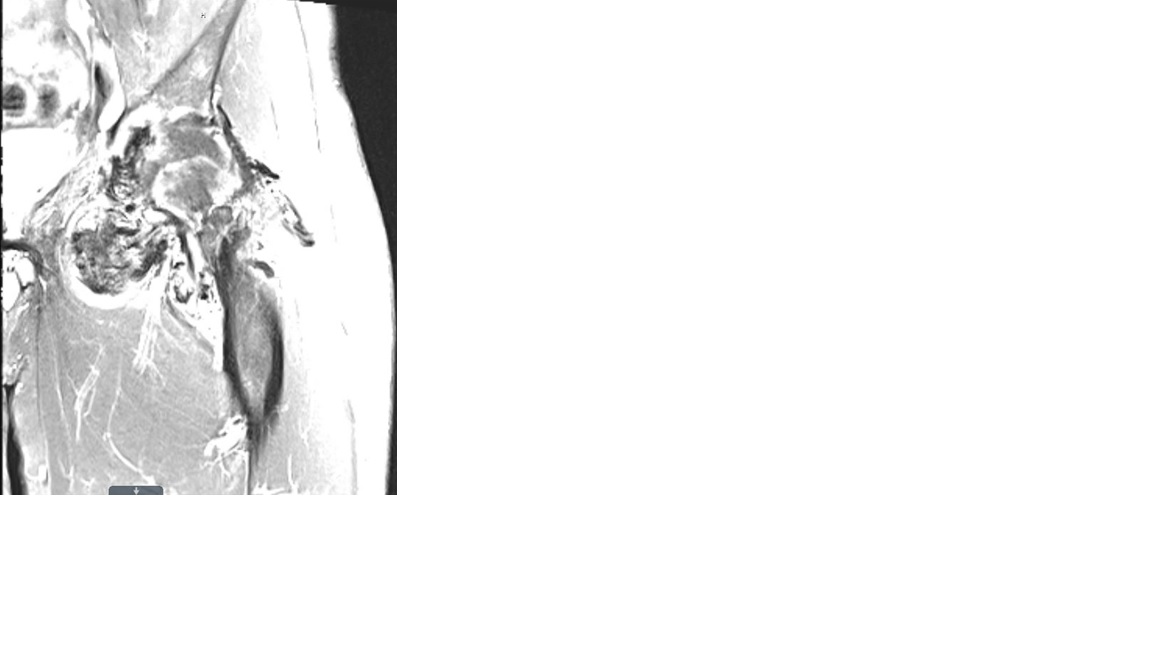
Case Presentation: A 49-year-old healthy man presented to the emergency department with a two-year history of left inguinal pain that had been increasing in frequency and severity, to the point where he experienced difficulty with ambulation. He denied fevers, chills, weight loss, weakness or numbness in the extremity. Physical exam revealed a palpable left groin mass. CT scan of the abdomen and pelvis demonstrated a 14.4 x 8.2 cm large soft tissue mass within the left hip and proximal left thigh musculature with associated mixed lytic and sclerotic osseous erosion of the left femoral neck, head, and acetabulum. Additionally, there were 3 subcentimeter hypo-attenuating hepatic lesions and an 8 mm lung nodule. These findings raised concern for a metastatic process such as sarcoma. The patient was admitted to the hospital medicine service for expedited evaluation and pain control. MRI of the hip showed pronounced enlargement of the left hip joint capsule with marked low signal internal contents on all sequences within the joint space. These findings were consistent with pigmented villonodular synovitis (PVNS). Core needle biopsy of the hip mass confirmed the diagnosis. PET scan revealed hypermetabolic large soft tissue mass within left hip and proximal thigh. No hepatic lesions were seen and the lung nodule was too small to be characterized. The case was discussed in tumor board and recommendation was for nonsurgical medical management with Imatinib or observation with repeat imaging in 6 months. The patient opted for observation and did not follow up in the clinic afterwards.
Discussion: PVNS is a rare, proliferative disease of the synovial joint, bursa, and tendon sheath which is often benign. Historically, pathogenesis of the disease thought to involve both inflammatory and neoplastic components. More recent studies have shown overexpression of colony stimulating factor 1 (CSF1) and favors neoplastic origin. PVNS has pathognomonic appearance on MRI, presenting with low signal intensity. Histologic confirmation remains the gold standard. Management strategy depends on type, size and aggressiveness of disease but is often surgical excision. For patients who are not surgical candidates or have recurrent disease, treatment with tyrosine kinase inhibitors, which have activity against CSF1 receptor, can be considered. Recently, Pexidartinib, a CSF1R inhibitor, has shown promising results in a randomized phase 3 clinical trial and now is been approved by FDA as a first systemic therapy for PVNS. Recurrence rates range from 10-15% for localized disease and up to 33% for diffuse disease.
Conclusions: We report this case of PVNS to increase hospitalists’ awareness about this benign but aggressive-appearing disease. Lack of knowledge about this rare disease could potentially lead to premature assumption of a malignant process. This case also highlights the importance of advanced imaging in the current era of medicine. Development of new targeted medical therapies can now offer a valuable treatment option for this debilitating condition, especially in unresectable cases.