Case Presentation: Cryptococcus is known for causing meningoencephalitis in immunocompromised individuals. We present a case of cryptococcal infection outside the central nervous system in a relatively immunocompetent host.
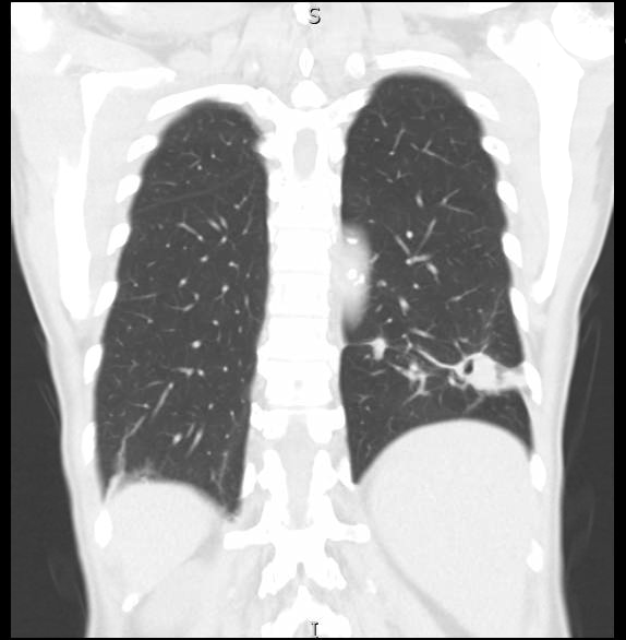
A 77 year-old male who immigrated from India at twenty years of age, with history of polycythemia vera, initially presented with hoarseness, occasional non-productive cough, weight loss and night sweats for two months. Of note, he is a non-smoker with no recollection of significant inhalational exposure. A chest x-ray (CXR) showed left lower lobe (LLL) consolidative changes. A subsequent computed tomography (CT) of the chest with contrast demonstrated a 2.4cm x 6.3cm nodular cavitary lesion within the LLL with adjacent smaller nodules within the parenchyma. A positron emission tomography (PET) scan correlated with a LLL fluorodeoxyglucose (FDG) avid mass. The working diagnosis at this time was malignancy. Approximately one month after, he underwent bronchoscopy with navigation – where brush and forceps biopsies and bronchoalveolar lavage (BAL) were performed. The results were negative for malignancy, contrary to the main diagnosis. Acid-fast bacillus smear was also negative. He was followed up with two serial CT chests done two months apart which showed no significant change. Finally, he underwent video assisted thoracoscopic surgery (VATS) with wedge resection of the LLL and chest tube placement. The postoperative course was complicated by heavy bleeding from the chest tube requiring surgical exploration and control of the bleeding. He was discharged on day five of admission.
Interestingly, the pathology revealed multiple necrotizing granulomata and focal emphysematous changes. The gram stain and mucicarmine stains were positive in yeast form consistent with Cryptococcus. Serum cryptococcal antigen sent subsequently, also returned positive at titers 1:8. Lumbar puncture was negative for central nervous system infection. The decision was made to place the patient on a six month course of fluconazole which is currently ongoing. Upon further interrogation, he endorsed living on a farm in India before emigration.
Discussion: Pulmonary cryptococcosis may have a wide range of clinical manifestation from asymptomatic to respiratory failure. Pulmonary nodules are the most common imaging finding associated with this diagnosis. Although there have been reported cases of pulmonary cryptococcosis, it remains relatively uncommon particularly in the immunocompetent patients. It is also a diagnosis which is likely to be missed with possible adverse sequelae. We may have spared this patient a partial lung resection with complications had cryptococcosis been diagnosed and treated earlier. Of note, as per the Infectious Disease Society of America guidelines one can choose to not treat localized pulmonary infections. However, this patient was treated due to the relatively high titres and history of polycythemia vera.
Conclusions: This case demonstrates the importance of taking a detailed history and having wide differential diagnoses- including rare causes such as Cryptococcus in a patient presenting with a lung nodule.