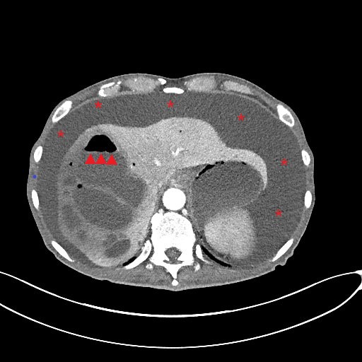
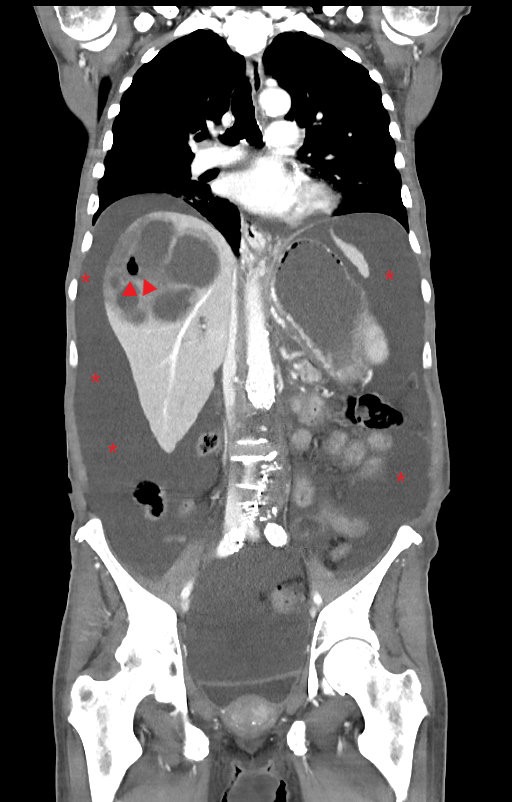
Case Presentation: A 70 year male presented to the emergency department with complaints of worsening fatigue, abdominal distension, abdominal pain, dyspnea, and increased lower extremity edema. He had been diagnosed with pancreatic cancer 3 months ago and recently started on chemotherapy. An abdominal CT scan revealed massive ascites and multiple liver abscesses essentially replacing the majority of the right hepatic lobe. The largest abscess measured 8 x 6 x 10 cm. The patient was treated with broad-spectrum IV antibiotics and percutaneous hepatic abscess catheter placement. Abscess fluid cultures were polymicrobial and included growth of Streptococcus constellatus, Escherichia coli, Prevotella buccae, Candida lusitaniae, and Slackia exigua. Unfortunately, the patient developed septic shock with bacteremia and multi-organ failure and died after six days of hospitalization.
Discussion: We describe a case of a patient with recently diagnosed pancreatic cancer who developed multiple, large pyogenic liver abscesses with striking radiologic findings. Of notable importance in this case, the abscesses developed rapidly within the span of 3 months, as a prior CT scan showed no liver abnormalities. The most common etiology of PLA is typically biliary tract disease, but other risk factors include hepatobiliary or pancreatic malignancy, liver transplant, diabetes mellitus, and use of proton pump inhibitors. In the last several decades, reports have shown an increase in prevalence of PLA in the US from 2.7 to 4.1 per 100,000 population.
Conclusions: Pyogenic liver abscesses are becoming more common in this country, especially those associated with malignancy. Patients with hepatobiliary or pancreatic malignancies are at increased risk due to biliary obstruction and possible immunosuppression from chemotherapy. This case serves as an impressive example of large multiple hepatic abscesses that can develop rapidly in the setting of pancreatic cancer, leading to a high risk of mortality.