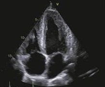
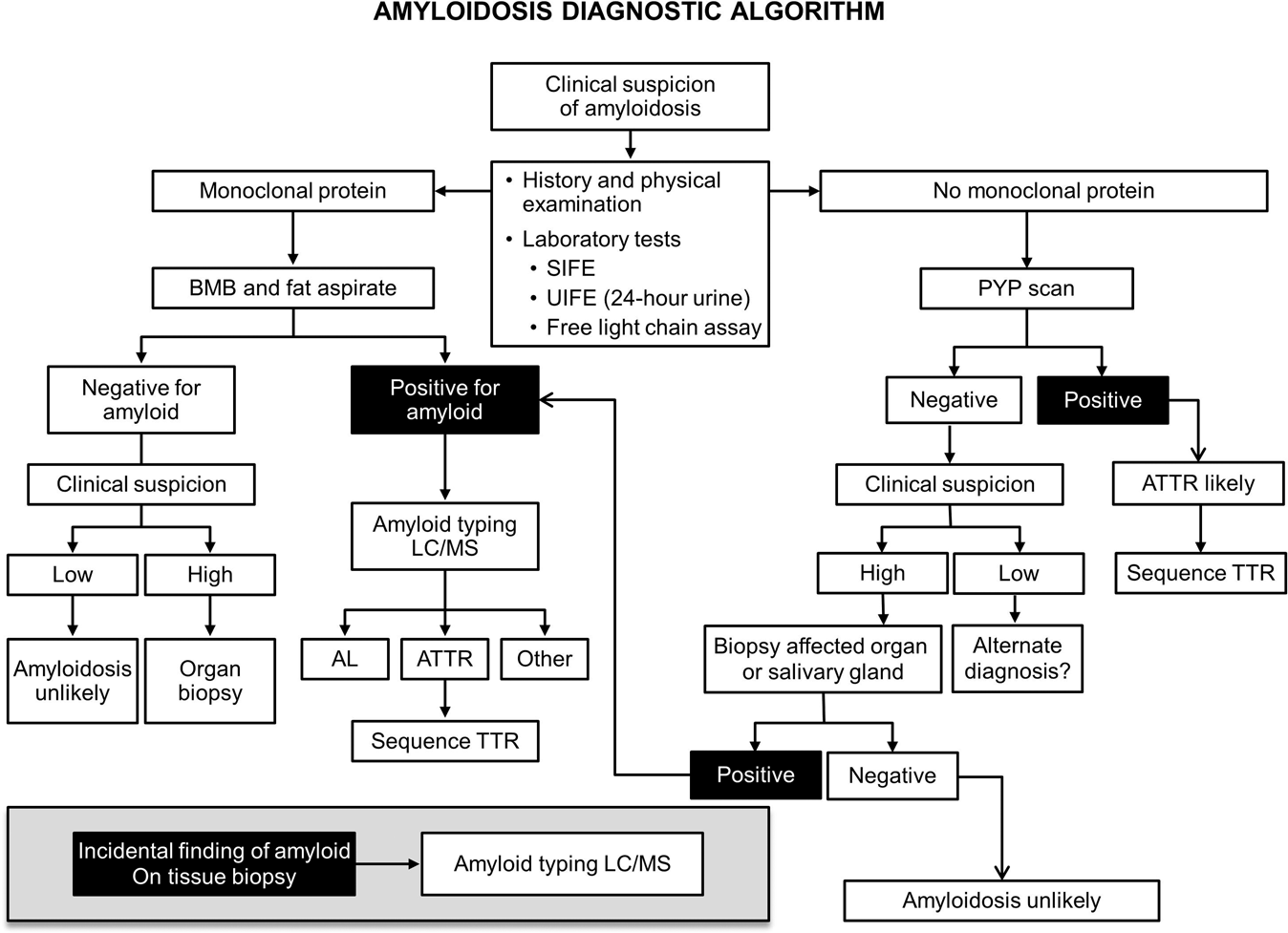
Case Presentation: A 70-year-old male presented with generalized fatigue and worsening shortness of breath with exertion. A diagnosis of acute exacerbation of heart failure was made. Initial evaluation with echocardiogram and MRI raised suspicion for amyloidosis. A PYP scan was performed, which was not suggestive for transthyretin amyloidosis (ATTR). Serum free light chain (sFLC) testing revealed a lambda monoclonal gammopathy with a differential light chain ratio (dLFC) of 125.8, free lambda levels of 160.3 mg/dL, and free kappa levels of 34.5 mg/dL. A monoclonal protein spike was detected in the serum.Endomyocardial biopsy confirmed the diagnosis of cardiac amyloidosis. Microscopic examination revealed mild myocyte hypertrophy, with perimyocyte and interstitial deposition of amorphous eosinophilic material that stained positive for amyloid. Immunohistochemical staining revealed faint perimyocytic lambda light chain staining, with no transthyretin detected.Bone marrow smear showed 1% plasma cells, and CD138 staining revealed 5-7% scattered plasma cells. Flow cytometry identified 0.8% lambda-restricted monoclonal plasma cells, as well as 0.2% kappa-predominant B cells expressing CD5, CD19, and CD20, which suggested the possibility of B cell lymphocytosis. The diagnosis of lambda light chain AL amyloidosis was confirmed, and the patient was started on treatment per the ANDROMEDA protocol with daratumumab, bortezomib, cyclophosphamide, and dexamethasone. Unfortunately, the patient passed away from cardiac arrest 21 days after the diagnosis.
Discussion: This case highlights the aggressive nature and diagnostic complexity of cardiac amyloidosis, specifically lambda light chain AL amyloidosis. Despite being a rare condition, amyloidosis should be considered in the differential diagnosis of heart failure, particularly in patients with preserved ejection fraction. Early diagnosis is critical, as delayed recognition often results in irreversible organ damage and poor outcomes.The patient’s negative PYP scan ruled out transthyretin amyloidosis (ATTR), while the positive serum free light chain assay and monoclonal gammopathy pointed towards AL amyloidosis. Endomyocardial biopsy confirmed the diagnosis, with histopathology revealing amyloid deposition and immunohistochemical staining identifying lambda light chain involvement. This diagnostic approach is crucial in distinguishing between AL and other types of amyloidosis, which have different prognostic and therapeutic implications.Although treatment was initiated according to the ANDROMEDA protocol, the patient succumbed to cardiac arrest shortly after diagnosis. This underscores the high mortality associated with cardiac AL amyloidosis, even when therapy is promptly started. Early detection through advanced imaging techniques, serum markers, and biopsy remains essential to improving survival.
Conclusions: This case also emphasizes the importance of comprehensive cardiac assessment in patients with suspected amyloidosis, as well as a multidisciplinary approach to management, including hematology, cardiology, and oncology input. Continued research is needed to improve outcomes, especially in the context of newer therapies targeting plasma cells and amyloid deposition. Nonetheless, even with current therapies, outcomes remain poor for patient was advanced cardiac amyloidosis. This underscores the need for continued advancements in both diagnostic tools and therapeutic options to improve survival.