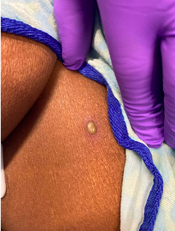
Case Presentation: A 60-year-old woman with past medical history of Systemic Lupus Erythematous (SLE) on daily mycophenolate mofetil and hydroxychloroquine, Antiphospholipid Syndrome on warfarin, history of aneurysm s/p clipping and diabetes presented to an urgent care clinic due to severe headache and dizziness. During examination, she was noted to have hesitancy with neck flexion, prompting her admission to the Emergency Department. Of note, she had previously been seen in urgent care the week prior for presumed viral pharyngitis with symptoms of sore throat, ear discomfort, and scattered pruritic bullae in a non-dermatomal distribution.On presentation, physical exam revealed stable vital signs, blurry vision with nystagmus upon looking to the left, and 4/5 strength in the left arm and leg. She had 10-15 non-pruritic and non-painful bullae, each approximately 1cm2 in size, scattered bilaterally on the shoulders, chest, arms, and legs. Labs were notable for an elevated International Normalized Ratio (INR) of 5.7. Imaging of the head including Computed Tomography (CT), CT Venography, and CT Angiography was negative for cerebral hemorrhage, venous sinus thrombosis, and cerebral arterial occlusion, respectively. Due to her supratherapeutic INR, a Lumbar Puncture (LP) was unable to be performed at the time of admission, and she was started on IV acyclovir for empiric viral meningitis coverage while Dermatology and Neurology consultations were performed. After negative imaging, improving neurologic symptoms, and consultative evaluation, there was a low clinical suspicion for disseminated Varicella Zoster Virus (VZV) prompting discontinuation of IV acyclovir.Dermatopathology subsequently returned suggestive of cutaneous herpetic infection, which was confirmed by skin lesion PCR positivity for VZV. After INR decreased, an LP was performed which found a Cerebrospinal Fluid (CSF) WBC of 55 that was 85% lymphocytic, CSF glucose of 97, and CSF protein of 45. CSF meningoencephalitis panel later resulted positive for VZV. IV acyclovir treatment was restarted, and the patient reported further rapid improvement of her headache and dizziness.
Discussion: Herpes Zoster affects more than 1.2 million adults annually in the United States (1), caused by reactivation of latent VZV in the sensory ganglia of the skin. This rash typically occurs in a unilateral dermatomal distribution. Reactivated VZV can disseminate widely in the body, particularly in immunocompromised hosts, causing complications such as meningoencephalitis, hepatitis, and pneumonia.This patient presented with an atypical neurologic and cutaneous presentation of disseminated Herpes Zoster infection, likely due to immunosuppression from her SLE medications. While extremely rare, a handful of cases of bullous VZV have been reported in both immunocompromised elderly and pediatric patients. In one case, a 69 year-old patient with past medical history of lymphoma, chronic kidney disease stage II, and type II diabetes was found to have a bullous rash with more than 20 lesions in the left C5-T1 dermatomes due to disseminated VZV (2).
Conclusions: Here we report an atypical case of disseminated Herpes Zoster. This case illustrates the importance of keeping a broad differential as atypical VZV infections can resemble other cutaneous conditions, muddying the clinical picture and potentially delaying prompt diagnosis and treatment.