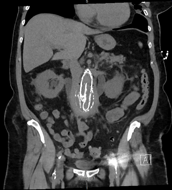
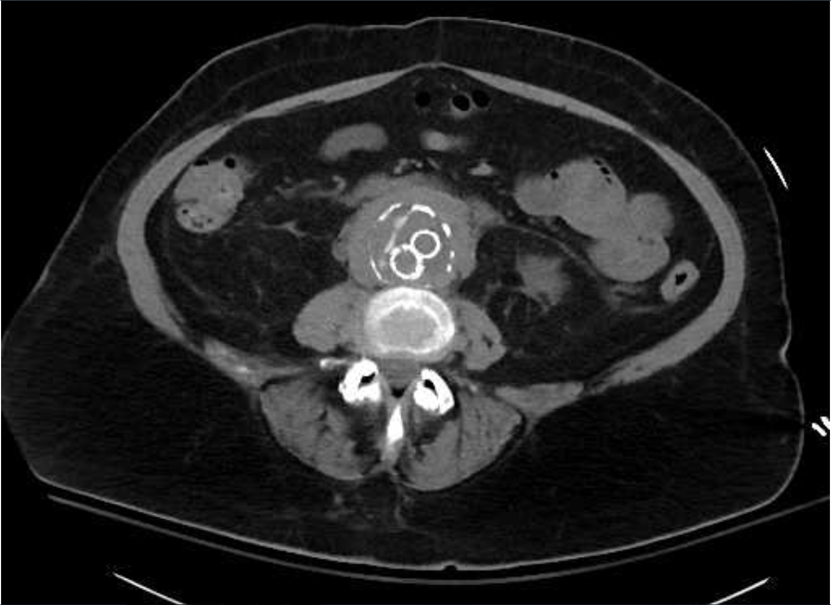
Case Presentation: Case 1: A 66 year old female presented in October 2017 with generalized weakness, fatigue, abdominal pain, and oliguria. The symptoms have been worsening since her abdominal endovascular aneurysm repair (EVAR). She had her EVAR done in August 2017. In the Emergency Room the patient was afebrile with a temperature of 36.4 C, pulse of 76 bpm, respiratory rate of 19, and blood pressure of 193/105. Laboratory studies showed a BUN of 98mg/dL, Creatinine of 17.93mg/dL, Sodium of 128mmol/L, Potassium of 6.1mmol/L, and Anion Gap of 18. A CT chest/abdomen/pelvis was done, and showed moderate bilateral hydroureteronephrosis, increased soft tissue thickening surrounding aneurysm sac suggestive of inflammatory/fibrotic changes and ureteral obstruction. A C-Reactive Protein level of 21.60mg/dL and a Erythrocyte Sedimentation Rate of 96mm/hr were found. She was admitted to the ICU and a Nephrology and Urology consult were placed. Bilateral ureteral stents were placed and the patient started to improve.
Case 2: A 70 year old male presented in November 2017 after being found to have acute abnormalities in his lab work found by Nephrology as an outpatient. He follows with Nephrology and Urology since having been diagnosed with retroperitoneal fibrosis and had bilateral ureteral stents placed. The patient’s Creatinine level was found to be 8.26mg/dL, with baseline around 3.0mg/dL. In the Emergency Room he was acidotic with a bicarbonate level of 13mmol/L, other lab results included a BUN level of 144mg/dL, Potassium level at 5.3mmol/L, Magnesium level of 1.0mg/dL. A bicarbonate drip was started. Nephrology and Urology were called and tried placing ureteral stents, but failed due to fibrotic tissue surrounding the ureters. Bilateral nephrostomy tubes were placed, and patient’s renal function initially started to improve, but started to decline again. He started showing signs of uremic encephalopathy, so a permacath was placed and he started hemodialysis. His renal function improved significantly. This patient was diagnosed with retroperitoneal fibrosis about three months status-post his EVAR.
Discussion: Primary immune-mediated processes are the more common cause of retroperitoneal fibrosis, but it is imperative to rule out secondary causes first. AAA surgery with EVAR has been increasingly more common due to the minimal invasive technique. Other complications of EVAR are endoleaks, graft thrombosis, and infection which all may have a component of an immunological mechanism causing the fibrosis. Treatment of this condition is typically steroids, and tamoxifen. Must avoid the use of medications that increase the risk of retroperitoneal fibrosis. It is important that this fibrotic process be caught early due to rapid progression with a high risk for acute renal failure requiring nephrostomy tubes and possible dialysis.
Conclusions: There have not been many reported cases of peri-aortitis and retroperitoneal fibrosis as a complication with EVAR. The few cases theorize that it may be due to local adventitial immune reaction to atherosclerotic plaque antigens and stent graft placement may facilitate the penetration of the plaques into deeper layers causing the inflammatory reaction. Patient’s may remain asymptomatic for a long period of time and so it is vital to continue to closely monitor the patient post-procedure as an outpatient. The most common presenting symptom is oliguria, this should immediately prompt a workup for obstruction – suspecting developing retroperitoneal fibrosis.