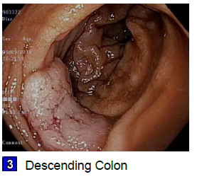
Case Presentation: A 59 year old female with a history of Hepatitis C cirrhosis, esophageal varices, type 2 diabetes mellitus, end-stage renal disease, and hypertension presented to the emergency department to be evaluated for fever at the behest of her dialysis center. She endorsed chills, diffuse abdominal pain, and dyspnea over the past week. She reported an episode of near-syncope two weeks prior to presentation, and had noted several episodes of hematochezia over the preceding three months which had been attributed to known oozing polyps. Upon physical examination, the patient was in no distress. Her blood pressure was 108/41, heart rate 91, temperature was 101.2 deg F, and respiratory rate 22 breaths per minutes, with oxygen saturation 94% on room air. She had bibasilar rales, abdominal distension and diffuse abdominal tenderness, and had a 3/6 systolic murmur at the left upper sternal border. Her digital rectal exam revealed no blood or mass. The patient’s white blood cell count was 8,800/mm3 with a neutrophilic predominance, hemoglobin was 8.4 g/dL, and platelet count was 67,000/mm3. Biochemistry results were in keeping with her baseline renal and hepatic disease. Bacterial blood cultures grew Strep sanguinis. Transthoracic echocardiography revealed a thickened aortic valve with new severe aortic regurgitation, which was noted to be concerning for infective endocarditis. A trans-esophageal echocardiogram was deferred due to her history of severe esophageal varices. Her severe periodontal disease was presumed to be the source of her bacteremia, and she had five teeth pulled prior to discharge home on six weeks intravenous Ceftriaxone. However, she came back to the emergency department two days after discharge with bright red blood per rectum. Colonoscopy performed the following day revealed a bleeding 3 cm partially circumferential non-obstructing mass in the descending colon. Pathology would reveal invasive adenocarcinoma. The Colorectal Surgery team determined she was not a surgical candidate due to Childs B cirrhosis and end stage renal disease. She is currently being considered for palliative radiation.
Discussion: The link between S bovis bacteremia and colorectal carcinoma has been well described. This case lends itself toward the growing body of evidence that viridans group strep species, like S sanguinis, should also prompt the physician to consider occult colonic malignancy. S sanguinis bacteremia as a harbinger of colorectal cancer has only been described 9 times in the literature so far. So far less than half of the referenced S sanguinis cases have been identified as having endocarditis. In this case we may presume that the patient had bacterial translocation from the gastrointestinal tract into the bloodstream. Since S. sanguinis is a common finding in oral flora, the physician may tend to attribute this blood stream infection to poor dentition, without pursuing a more invasive workup, as was the case for our patient. Her colonoscopy was therefore delayed until she represented with hematochezia.
Conclusions: Thorough workup is required in cases of unexplained bacteremia involving Strep sanguinis, similar to that which is routine in Strep bovis. Caution should be exercised in attributing Strep sanguinis bacteremia to poor dentition without further workup. Further studies are needed to ellucidate the frequency of S sanguinis blood stream infections heralding colonic malignancy.