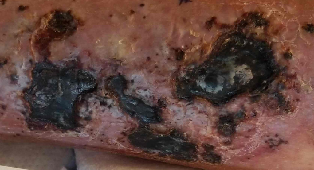
Case Presentation: 44 year-old male with ESRD on HD complicated by tertiary hyperparathyroidism, possible calciphylaxis on treatment with sodium thiosulfate, possible adrenal insufficiency on steroids, antithrombin III deficiency with prior pulmonary and splenic infarcts on Coumadin who developed a diffuse cutaneous skin eruption during a recent admission after being on antibiotics for possible pneumonia. He was discharged to a SNF and presented two weeks later with hypotension and worsening rash. On exam, patient was found to have erythematous macules involving all four extremities including the palms, trunk and back which then became erupting vesicles that formed necrotic eschars over his extremities. A biopsy revealed small vessel leukocystoclastic vasculitis. Given the degree of his rash, he was transferred to our hospital for further management. On exam he was found to have retiform necrotic ulcerations with thick eschars on his extremities, as well as numerous purpuric macules coalescing into pat ches. Sheets of firm subcutaneous plaques were noted of the bilateral medial thighs and lower abdomen. APLS and cryoglobulins were negative. PTH and calcium were elevated. Xray imaging of his lower extremities showed ossification and foci with rim-like mineralization. Dermatology and Rheumatology were consulted and felt patient had leukocytoclasic vasculitis in the setting of calciphylaxis. Diagnosis of calciphylaxis was based on subcutaneous plaques consistent with calcium deposition and the xray imaging showing calcium deposition. They recommended tacrolimus cream for his vasculitis, as well as continued treatment with sodium thiosulfate for his calciphylaxis. Further, dermatology recommended to switch patient to apixaban.
Discussion: This case is relevant to internal medicine physicians as it demonstrates the importance of diagnosing calciphylaxis, and identifying risk factors that may contribute to this disease process. Calciphylaxis is characterized by calcification and occlusion of the microvasculature in the subcutaneous adipose and dermal tissue, and is most commonly seen in patients with ESRD. Classic presentation is of intensely painful skin lesions which appear as plaques, nodules, livedo or purpura which rapidly progress to necrotic ulcers with eschars, similar to our patient’s presentation. There are a many risk factors associated with calciphylaxis including obesity, DM, hypercalcemia, hyperparathyroidism, hypercoagulable states, autoimmune disorders, and medications including corticosteroids, coumadin, and heparin. A diagnosis can be presumed in a patient with ESRD who presents with necrotic ulcers and palpable subcutaneous plaques. Treatment focuses on eliminating risk factors, as in our patient. His Coumadin and steroids were stopped, and his tertiary hyperparathyroidism was treated with cinacalcet and sodium thiosulfate. Despite addressing modifiable risk factors and treating with sodium thiosulfate, calciphylaxis ultimately carries a poor prognosis.
Conclusions: Calciphylaxis is a feared complication of ESRD that is associated with extremely poor outcomes. It is a rare and challenging diagnosis to make but if suspected efforts should be made to avoid worsening the condition.