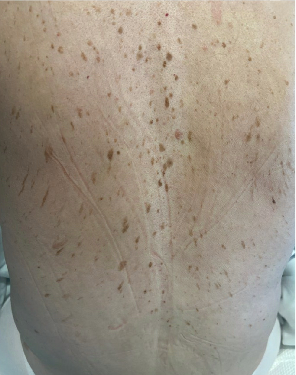
Case Presentation: A healthy 64-year-old woman presented with a sudden onset of crampy lower abdominal pain, nausea, and vomiting. Physical examination showed a soft, distended abdomen with lower abdominal tenderness and multiple waxy, brown to tan colored papules and plaques on the back, resembling “raindrops,” consistent with seborrheic keratoses. This skin finding was new; her previous evaluation a year ago had shown only a few scattered seborrheic keratoses. A CT scan of the abdomen and pelvis revealed a left adnexal mass, suggesting an ovarian neoplasm. Tumor markers CA 19-9, CA 125, and hCG were elevated, while CEA levels were normal. Colonoscopy and EGD revealed a few hyperplastic polyps in the colon and gastric polyps in the stomach, but biopsies were negative for malignancy. The patient underwent diagnostic laparoscopy followed by exploratory laparotomy and extensive debulking of the ovarian tumor. The pathological analysis confirmed a mixed carcinoma, predominantly clear cell carcinoma (80%) with a minor component of endometrioid carcinoma (20%). The patient is currently undergoing adjuvant chemotherapy.
Discussion: The Leser-Trélat sign, characterized by the abrupt emergence of multiple seborrheic keratoses, has long been recognized as a paraneoplastic cutaneous marker of internal malignancy. Common malignancies associated with this sign are gastrointestinal adenocarcinomas (gastric, colon, rectal), out of which gastric adenocarcinoma is the most common. It is proposed that the underlying malignancy triggers the overproduction of growth factors such as Epidermal Growth Factor alpha (EGFα) and Epidermal Growth Factor Receptor (EGFR), leading to the formation of seborrheic keratosis lesions. The Leser-Trélat sign can manifest before, during, or after the diagnosis of malignancy. High clinical vigilance is needed when evaluating patients presenting with this sign, as it can often be mistaken for benign seborrheic keratoses, especially in the elderly population. A key differentiator is the rapid onset of seborrheic keratoses or a significant increase in the number of lesions in a short timeframe, frequently accompanied by unusual pruritus, which should raise suspicion of an underlying carcinoma. Furthermore, this sign’s appearance in a younger demographic may be a stronger indicator of an associated malignancy, as seborrheic keratosis is less common in younger patients. With the disappearance of the primary tumor, paraneoplastic dermatoses often resolve, but may reappear with tumor recurrence.
Conclusions: This case demonstrates an unusual occurrence of the Leser-Trélat sign in the context of ovarian cancer, expanding the spectrum of this paraneoplastic manifestation. Moreover, amidst advancements in medical technology and diagnostic tools, this case highlights the importance of conducting a thorough physical examination as early recognition can facilitate prompt diagnosis and timely management of the associated malignancy.