Case Presentation: 51yoM with no significant medical history presents to ED for evaluation of progressive bilaterally injected, painful conjunctiva of 1 months duration, nonproductive cough and pleuritic chest pain of 1 week’s duration. He was recently treated with moxifloxacin for conjunctivitis without improvement in symptoms. After treatment, he developed nonproductive cough associated with pleurisy and post tussive emesis. In the ED, vitals were stable and WNL.
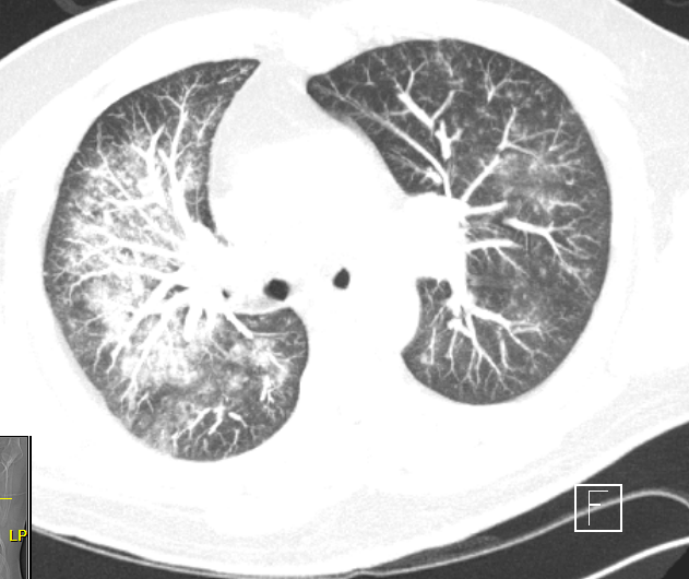
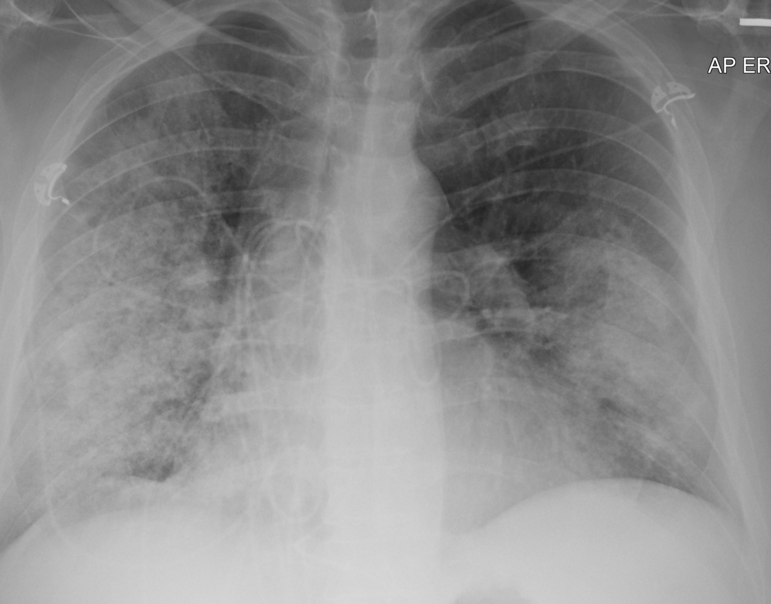
Labs were notable for mild hypovolemic hyponatremia, hypokalemia leukocytosis (10.1), with 77% neutrophils, elevated CRP of 189.6. EKG and serial troponins were WNL. Chest x-ray revealed bilateral mid and lower lung field airspace opacities. Follow-up CT chest with angiography revealed bilateral confluent ground-glass and interstitial opacities with peripheral sparing. He was admitted for the management of pneumonia versus pneumonitis. Ophthalmology was consulted. After evaluation, patient was diagnosed with superficial punctate keratitis and was prescribed tobramycin/dexamethasone ophthalmic solution TID for 1 week.
During his hospitalization, he became febrile and hypoxic. He subsequently underwent bronchoscopy with BAL which revealed serial aliquots concerning for alveolar hemorrhage. Unfortunately, following bronchoscopy his respiratory status deteriorated and patient required intubation. Out of concern for DAH, as result of vasculitis C-ANCA, P-ANCA and complement were ordered.
He had positive C ANCA 1:4096, positive PR3 and normal complement levels. He was treated with high dose steroids and 5 sessions of plasmapheresis. Upon completion of plasmapheresis, weakly infusion of rituximab was initiated.
Discussion: Superficial punctate keratitis is nonspecific and relatively uncommon diagnosis of unclear etiology. There is no known association between SPK, DAH and GPA. Although granulomatosis with polyangiitis can be associated with ocular manifestations they are usually in the form of peripheral ulcerative keratitis and uveitis. Due to AR unspecific symptoms and hospitalization, there was high early suspicion for autoimmune disorder. This led to early testing for TB and hepatitis which shortened hospitalization.
Conclusions: Superficial punctate keratitis is relatively uncommon ocular disorder that requires slit tab examination for diagnosis. Clinically, it is defined by redness, tearing nose and blurry vision. It is often treated with ophthalmic cyclosporine or steroids. Its pathophysiology is poorly understood, but connective tissue disorders and vasculitis have been hypothesized to be contributing factors. When this disorder is suspected in an individual requiring inpatient treatment, it is important to consider autoimmune disorders as part of the patient’s differential. In our case with Mr. AR, his superficial punctate keratitis preceded his diagnosis of granulomatosis with polyangiitis and life threatening diffuse alveolar hemorrhage. With Mr. AR’s constellation of symptoms and ocular involvement, suspicion for vasculitis and autoimmune process was raised early and hepatitis serologies and QuantiFERON-TB Gold testing was ordered early. This allowed for expedited treatment of granulomatosis with polyangiitis once diagnosed and reduced length of hospitalization.