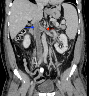
Case Presentation: A 56-year-old-male with hypertension, diabetes mellitus, chronic kidney disease (CKD) stage 3 presented with a 6-month history of intermittent lower extremity swelling and a 3-week history of scrotal and abdominal swelling. 9 months prior, he was noted to have worsening renal function and nephrotic range proteinuria (13g) prompting two renal biopsies which demonstrated diabetic nodular glomerulosclerosis and severe vascular sclerosis. At that time his hemoglobin (Hb) was 17 (13-18g/dL). On presentation, vitals were unremarkable. . Physical examination was significant for splenomegaly and bilateral lower extremity pitting edema extending to the scrotum associated with bilateral calf tenderness. Biochemical investigations were significant for Hb of 17, hematocrit of 51 (40-52%), platelet of 393 (150-400k/uL), albumin of 4.0( 3.5-5g/dL) and creatinine (Cr) of 2.7 (Baseline 1.8 (0.7-1.3mg/dL)). Venous doppler revealed bilateral acute occlusive deep vein thromboses in the external iliac vein, common femoral and femoral vein and popliteal veins. CT angiogram revealed thrombus involving the intrahepatic and intrarenal inferior vena cava (IVC), bilateral renal and gonadal veins. He was treated with intravenous heparin infusion, subsequent mechanical thrombectomy of the IVC, bilateral femoral and iliac veins, angioplasty and stenting of the proximal IVC. Post hydration, Cr improved to 1.4. Further investigations revealed an elevated LDH 827 (316-618 U/L), erythropoietin 3.7 (2.6-18.5 MIU/ML) and positive JAK2 V617f mutation. Antiphospholipid antibodies, C3/C4, rheumatoid factor and ANA were negative. His edema improved and he was discharged on Apixaban for long term anticoagulation and followed by hematology for polycythemia vera (PV).
Discussion: The prevalence of anemia increases with worsening CKD, from 8.4% at stage 1 to 53.4% at stage 5 (1). A high or normal Hb in a patient with CKD must be evaluated, especially when associated with thrombosis or worsening renal function as additional diagnoses may influence management, disease progression and mortality. PV is a myeloproliferative disorder (MPD) defined by the WHO criteria by Hb >16.5 in men, BM hypercellularity for age with trilineage growth, JAK2 V617F or exon12 mutation and low EPO levels (2). Hyper viscosity of blood in PV can cause thrombosis and worsening renal function (3). Although nephrotic syndrome is well known to increase the risk of thromboembolic events, patients such as in this case, presenting with elevated Hb and advanced CKD, splenomegaly and now extensive thrombosis, should prompt suspicion for a MPD. The worsening renal function in this case was likely secondary to a combination on DM nephropathy, dehydration and concurrent new renal vein thrombosis.
Conclusions: In patients with CKD, workup for other underlying causes of elevated Hb or hematocrit is advisable especially in the presence of advancing renal failure. Prompt recognition of MPDs may result in the early identification of additional risk factors for thrombosis, improving overall patient morbidity and mortality.