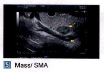
Case Presentation: A 65 year-old asymptomatic female was referred to a gastroenterology office for a new diagnosis of Hepatitis C. Labs were remarkable for mildly elevated ALT (49) and AST (53). A routine computed tomography (CT) of abdomen/pelvis revealed a 2.3 cm x 2.1 cm x 3.1 cm well circumscribed, enhancing, and hypovascular mass in the uncinate process of the pancreas. Subsequently, an endoscopic ultrasound (EUS) with biopsy was performed and pathology was positive for S-100 spindle cell neoplasm consistent with nerve sheath tumor. Patient underwent pylorus preserving Whipple’s procedure without any postoperative complications and was discharged with an unremarkable course afterwards.
Discussion: Schwannomas are mesenchymal neoplasms originating from the Schwann cells of the nerve sheath. In general, they are benign, encapsulated, and slow growing; however, approximately 10% have malignant potential. Pancreatic schwannomas are very rare. The average age at diagnosis is 55 with females (57%) having slightly higher incidence than males (43%). About one-third of the patients are asymptomatic while the rest experience non-specific symptoms including abdominal pain, nausea, vomiting, dyspepsia and back pain. No specific tumor markers are associated with them. Diagnosing schwannoma is often challenging on imaging, such as CT, ultrasound, and MRI, as it mimics other cystic tumors, like pancreatic pseudocysts, intraductal mucinous-papillary neoplasm, or serous/mucinous cystic neoplasms. Hence, they have a high rate of preoperative misdiagnosis. EUS with fine needle aspiration may aid in the definite preoperative diagnosis; however; there is a chance of high false negative. Surgery with histological examination and immunohistochemical staining (S-100 positive) is needed for definite diagnosis.
Conclusions: Pancreatic schwannomas are not only rare but are a great imitator as well. With non-specific clinical manifestation, lack of distinct characteristic findings on multiple imaging modalities, and high rates of false negatives on FNA, preoperative diagnosis is often difficult. These tumors demand special consideration with broader differential diagnoses since these tumors mimic a multitude of other neoplasms. Surgery is not only diagnostic but also therapeutic with patients generally having a good prognosis after tumor resection.