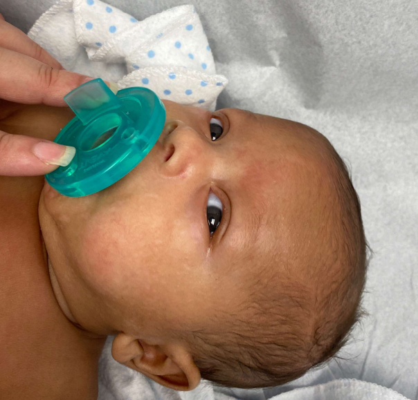
Case Presentation: An exclusively breastfed 2-month-old male born at 31 weeks gestation presented with 10 days of progressively worsening rash over his face, genitalia, and right upper extremity. Prior to transfer, the patient had previously been treated for suspected impetigo with topical mupirocin, oral cephalexin, and IV cefazolin without improvement. Upon presentation, the patient was alert and irritable but consolable with stable vital signs for age. His skin exam revealed diffuse erythematous, eroded papules and plaques, including some overlaid with yellow crusting, in the periocular area, bilateral cheeks, anterior and posterior neck, right forearm, inguinal folds, inferior penile shaft, foreskin, and perianal area. The remainder of the exam was normal.Labs demonstrated an ANC of 0.5 and a CRP of 1.3; negative GAS and aerobic cultures, HSV/VZR and enterovirus PCR antigen tests; and normal Vitamin A, B3, and D levels. His newborn screen was normal. His zinc level was low (31 ug/dL) and skin biopsy revealed epidermal hyperplasia with pallor in the upper epidermis and parakeratosis, highly suggestive of acrodermatitis enteropathica (AE). He was treated with oral zinc supplementation with improvement in the rash within a few days and complete resolution 2 weeks later. After his follow-up, his vitamin B7 (biotin) level resulted and was undetectable.
Discussion: Many newborn rashes are benign and self-resolving and do not require diagnostic testing or treatment. The age of the patient, characteristics of the rash, and associated symptoms are important when determining the need for further evaluation. This patient presented with well-demarcated, scaly, erythematous plaques in a periorificial distribution, with associated diarrhea and irritability. These findings, along with minimal response to antibiotic therapy, initially suggested AE. AE is classically caused by an autosomal recessive mutation in the infant’s zinc transporter gene SLC39A4, leading to improper enteral zinc absorption. AE typically manifests when infants are weaned from breast milk or earlier in infants who are formula-fed, neither of which occurred for this patient. Clinical response to zinc supplementation can be dramatic and occurs within a few days, as did this patient. Although this patient responded to zinc supplementation, a diagnosis of acrodermatitis dysmetabolica (AD) secondary to biotinidase deficiency is more likely. AD can be caused by an acquired zinc deficiency or other metabolic disorders unrelated to zinc. Biotinidase deficiency is associated with reduced zinc levels in animal models and presents with similar characteristic skin lesions. Although often detected on the newborn screen, infants with concerning clinical presentations or laboratory studies should undergo single-gene testing for BTD and biotinidase activity. Additional vitamin deficiencies to consider include Vitamin B2 (riboflavin) and fatty acids. This patient’s genetic testing is pending.
Conclusions: Scaly, erythematous periorificial plaques on the face and anogenital region should trigger both an infectious and nutritional deficiency evaluation given overlap in presentation.