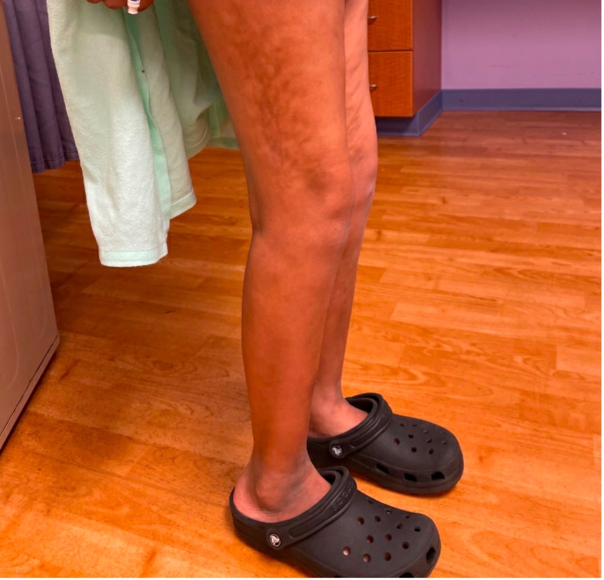
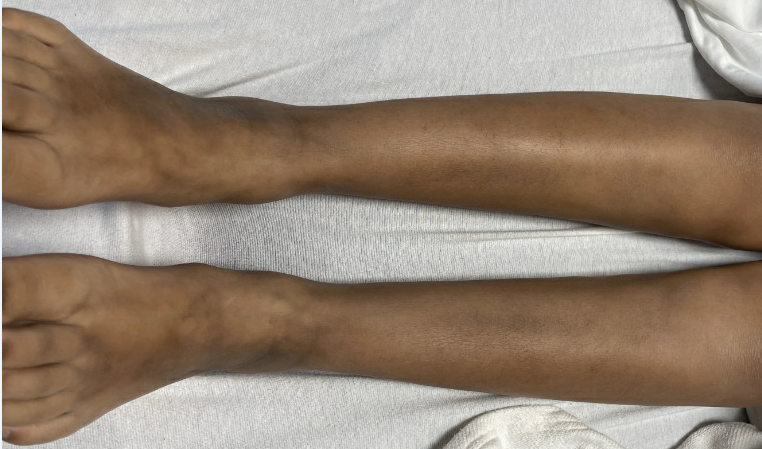
Case Presentation: A 9-year-old female presented with two months of hyperpigmented skin and stiffness mainly in her lower extremities. She had difficulty brushing her hair and walking accompanied by weight loss and dyspnea. On examination, diffuse hyperpigmentation and peau d’orange skin changes were observed with the “groove sign” on her right medial thigh. She also had restricted range of motion in her extremities affecting her gait. Laboratory results were significant for elevated eosinophils and inflammatory markers with negative antinuclear and scleroderma antibodies. A chest CT revealed fibrotic strands and interstitial thickening in both lungs, and severe restrictive lung disease was noted on pulmonary function tests. Subsequently, an MRI showed T2 hyperintensity in the subcutaneous tissues and muscles in her abdomen and upper thighs, but there was no fascial enhancement to suggest fasciitis. Ultimately, a muscle biopsy confirmed the diagnosis of eosinophilic fasciitis (EF). She received steroids and methotrexate followed by rituximab. Unfortunately, her disease continued to progress despite these therapies, and her course was complicated by posterior reversible encephalopathy syndrome and adrenal insufficiency. She has since improved with steroids, infliximab, and mycophenolate mofetil.
Discussion: EF is a connective tissue disorder characterized by progressive thickening of the skin and soft tissues. There are 300 cases documented to date with only 21 involving pediatric patients. Steroids is the mainstay of treatment providing resolution in most patients, while methotrexate, tumor necrosis factor-alpha inhibitors, and rituximab are reserved for more refractory conditions. EF is considered a “scleroderma mimicker” due to shared skin changes, but it is crucial to delineate the two diseases. Unlike scleroderma, EF is characterized by eosinophilic infiltration that primarily affects the skin, fascia, and muscles of the limbs in the absence of Raynaud’s phenomenon and nailfold capillary changes. Visceral involvement is rare, rendering this case unique, as pulmonary manifestations have been documented in fewer than five cases. Finally, a hallmark feature of EF is the “groove sign,” defined by linear depressions along superficial veins due to tissue fibrosis.
Conclusions: This case highlights a pediatric patient with refractory EF complicated by unexpected severe lung disease. EF is an inflammatory condition that causes scleroderma-like skin changes in the limbs, resulting in chronic debility. The prognosis is excellent in adults, but this disease can be more challenging to treat in children. A full-thickness muscle biopsy is the gold standard for diagnosis that can differentiate EF from scleroderma, allowing for timely initiation of therapy.