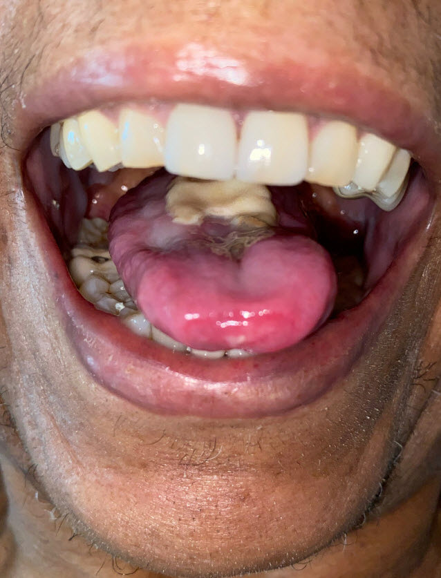
Case Presentation: A 73 year old female with a 50 pack year smoking history was admitted with 2 months of dysphonia, oral pain, odynophagia, and a 30 pound weight loss. Exam revealed a large ulcerative lesion of the tongue and white plaques on the tongue and oral mucosa. CRP was 5.74 mg/dL and ESR was 64 mm/hr. ENT, ID, dentistry, and rheumatology were consulted. Laryngoscopy and CT neck were normal. CT Chest showed patulous esophagus with distal esophageal wall thickening and scattered sub-centimeter pulmonary nodules. Biopsy of the tongue lesion showed acute inflammation with bacterial colonization and was negative for dysplasia, carcinoma, granulomas, vasculitis, HSV, VZV, CMV, fungal stain and culture. She was treated with fluconazole for oral and esophageal candidiasis with improvement of odynophagia. She was discharged with outpatient follow-up. Two months later, the patient was readmitted with acute onset blindness of her left eye and decreased vision in her right eye. She reported new-onset left-sided frontal headache associated with left eye pain and jaw fatigue with chewing. CRP was 1.93 mg/dL and ESR was 46 mm/hr. CBC was notable for leukopenia. Rheumatology and ophthalmology were consulted and performed a temporal artery biopsy. She was treated with methylprednisolone 1 gram for 3 days. The biopsy returned with evidence of arteritis but did not reveal any giant cells. MRI showed abnormal enhancement of the bilateral optic nerves and abnormal T2 signal consistent with optic neuritis. She was diagnosed with atypical giant cell arteritis. She was discharged on prednisone and subsequently started tocilizumab as an outpatient. She has not regained vision in her left eye but vision in her right eye improved significantly to 20/50.
Discussion: Giant cell arteritis (GCA) is a medium to large vessel vasculitis which classically presents as headache, vision changes, and jaw claudication.1 The American College of Rheumatology includes meeting three of five criteria for the diagnosis of GCA: age ≥ 50 years old, new onset of localized headache, temporal artery (TA) tenderness or decreased pulse, ESR ≥ 50 mm/hr, and positive TA biopsy. TA biopsy is the gold standard for GCA diagnosis.2 GCA can lead to devastating complications such as blindness and mortality, which is why early recognition and treatment is important. The differential diagnosis for tongue ulceration is broad, including trauma, malignancy, infection, or drug reaction. Given the large blood supply to the tongue, ulceration or necrosis of the tongue as a sole initial manifestation of GCA is exceedingly rare.3-5 In prior case reports of tongue involvement with GCA, most patients had other classic symptoms, such as headache, vision changes, and/or diminished temporal arterial pulse,6-11 none of which were present in our patient on initial presentation. Case reports have noted concomitant candidiasis in GCA, but there were other manifestations of GCA present to support the diagnosis.10,12 Biopsy of tongue lesions may be non-specific and not show signs of vasculitis13,14, so diagnosis relies on clinical recognition and pursuit of a TA biopsy.
Conclusions: Tongue ulceration is a rare manifestation of GCA. In this case, tongue ulceration preceded headache, vision loss, and jaw claudication by several months. GCA should be included in the differential diagnosis of tongue ulceration. Early diagnosis along with a high index of clinical suspicion is paramount to halting progression of this disease.