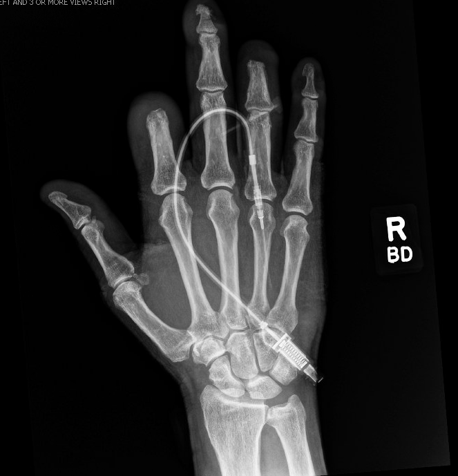
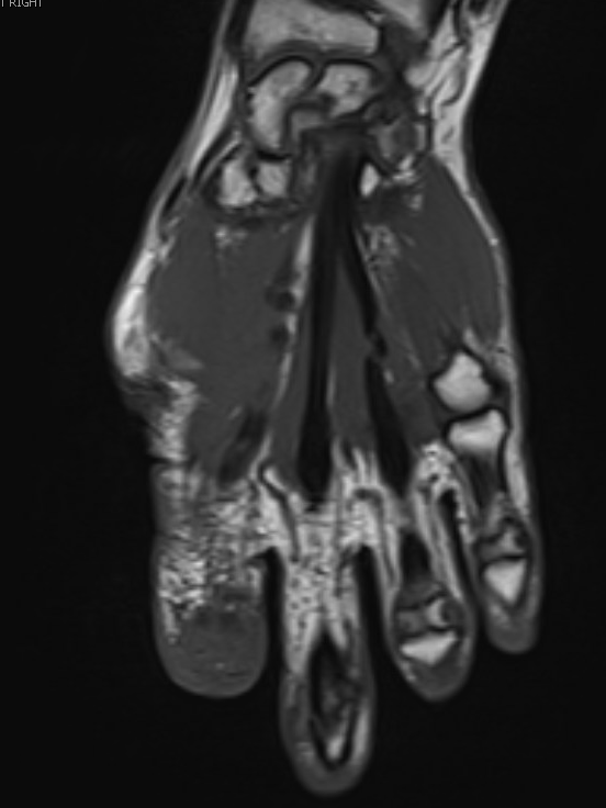
Case Presentation: A 61-year-old man with type 2 diabetes, autonomic neuropathy, prior right partial hallux amputation, right third metatarsal head resection, and previous right ring finger I&D presented with two weeks of swelling, erythema, drainage, and distal numbness of the right second finger. He has a longstanding finger chewing habit and had recently completed a 10 day antibiotic course for a UTI. Exam showed fusiform swelling and distal necrosis of the right second digit with preserved PIP/MCP motion; the left third distal digit also had necrotic tissue. Imaging demonstrated multifocal destruction of the right second distal phalanx and erosive changes of the left third digit consistent with osteomyelitis. He was started on vancomycin and piperacillin-tazobactam and underwent partial amputation of the right index finger; cultures grew MSSA, and he was transitioned to cefazolin with planned cephalexin.He was recalled when an AFB culture from the surgical specimen grew Mycobacterium tuberculosis. Pathology showed necrosis, granulation tissue, inflammation, cellulitis, and acute osteomyelitis. He denied TB exposures but reported 56 lb weight loss. Vitals were stable. Labs showed WBC 9.1, A1c 6.8, ESR 89, and CRP 21.3. Quantiferon, HIV, hepatitis B/C, MTB PCR, and blood cultures were negative; three sputum AFB smears and cultures were also negative. CT chest showed 2 small left lower-lobe nodules, and MRI revealed cellulitis of digits two–five with nonspecific marrow edema.He was admitted for tuberculous dactylitis and evaluation for pulmonary involvement. Airborne isolation, CT chest/abdomen/pelvis, inflammatory markers, serologies, and sputum studies were obtained. Psychiatry evaluation for finger-chewing was advised but declined; olanzapine was started. After three negative smears and PCR, isolation was discontinued. Infectious diseases recommended isoniazid, rifampin, pyrazinamide, ethambutol, and pyridoxine for 6–12 months with LFT monitoring. Because imaging suggested osteomyelitis and MSSA was levofloxacin-sensitive, he declined IV therapy and agreed to 6 weeks of oral levofloxacin. He was discharged with close follow-up and instructed to begin TB therapy under Department of Health supervision.
Discussion: Tuberculous dactylitis is a rare form of skeletal TB, typically seen in children and often absent of pulmonary findings. Adult cases are uncommon and easily mistaken for bacterial osteomyelitis because the presentation is indolent and routine tests IGRA, AFB smear, and PCR may be negative. This patient’s necrosis, MSSA positive cultures, and chronic finger chewing habit reinforced the initial bacterial diagnosis, delaying recognition of TB. The diagnosis was confirmed only through culture from surgical tissue. His bilateral digital involvement, weight loss, diabetes, neuropathy, and absence of exposures contributed to a complex, atypical presentation. Standard therapy includes multidrug anti-TB treatment for 6–12 months; surgery is usually diagnostic or reserved for severe destruction. He additionally required oral levofloxacin for MSSA associated osteomyelitis.
Conclusions: Adult-onset tuberculous dactylitis can closely mimic bacterial osteomyelitis, leading to delayed diagnosis and unnecessary surgery. Negative IGRA, AFB smears, and PCR do not rule out extrapulmonary TB; culture and biopsy remain essential. TB should be considered in chronic or recurrent digital infections that do not follow an expected clinical course.