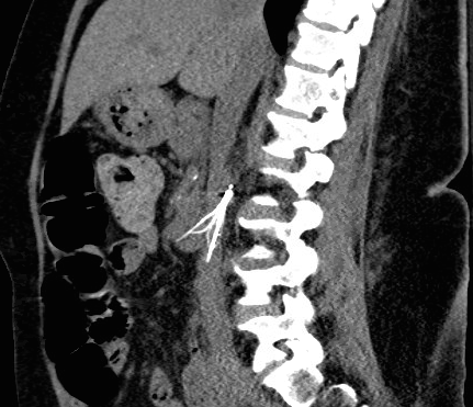
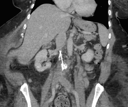
Case Presentation: A 54 year-old woman with history of pulmonary embolism and deep vein thrombosis (with prior placement of inferior vena cava (IVC) filter on apixaban) presents with recurrence of hematemesis and dark stool. This was her third hospitalization with GI bleed requiring transfusion in the last few weeks. Her prior work up included multiple negative studies, including colonoscopy, upper endoscopy, capsule study, and tagged red blood cell scan. One week into her admission, she had large volume hematemesis and hypotension requiring massive transfusion and ICU transfer. Endoscopy with push enteroscopy was performed with no source or active bleeding noted. She was stabilized and transferred back to the medical floor but she continued to have recurrent bleeds. A CT angiography was performed that did not identify active bleeding, but showed that the IVC filter was displaced, with prongs extending outside the lumen of the IVC and damaging nearby structures. One prong damaged the wall of the aorta, creating 1.3 cm pseudoaneurysm abutting the wall of the duodenum. A second prong penetrated the wall of the duodenum. This resulted in an aortoenteric fistula that was the source of ongoing bleedingShe subsequently underwent emergent surgery including repair of pseudoaneurysm, duodenal resection, and filter removal. Surgery was complicated by hemorrhagic shock from pseudoaneurysm rupture, resulting in cardiac arrest. Despite the complex surgery and intra-operative complications, she made an impressive recovery and was discharged to a rehabilitation facility.
Discussion: IVC filter remains controversial in the management of VTE. The only consensus indication for filter placement is in the setting of contraindications to anticoagulation (AC), AC complications, or failure of AC in setting of proximal DVT. Current CHEST guidelines recommend against the use of an IVC filter (Grade 1B) in patients with acute DVT or PE on AC. However, the indication and use of filters have expanded beyond these consensus recommendations, despite lack of evidence of efficacy and safety. The PREPIC trial showed that with placement of a permanent IVC filter, any benefit of reduced PE was masked by the increased proximal DVT, and with no apparent effect on overall VTE incidence or mortality. This was followed by the PREPIC 2 trial that found that placement of a retrievable IVC filter for 3 months did not reduce recurrent or fatal PE. Other studies measuring removal of retrievable filters suggest the retrieval rate is much lower (on the order of 10-20 percent) outside the ideal conditions of PREPIC 2 (where 80 percent were retrieved). The low rate of retrieval is significant given the complications associated with long-term filter retention, including migration and damage to nearby structures such as the IVC, aorta, and heart. These complications were highlighted in 2010 and 2014 FDA safety communications. It is recommended to remove retrievable filters in as little as several weeks after placement to reduce complications associated with retention or migration.
Conclusions: This case highlights a rare but serious complication of a retained IVC filter, aorto-enteric fistula, which is an an elusive cause of massive GI bleed. The patient had recurrent life threatening hemorrhage due to filter breakage and migration, requiring emergent high-risk surgical repair. Limiting IVC filter placement to appropriate indications and ensuring timely followup for removal can reduce the risk of complications associated with retained filters.