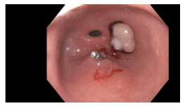
Case Presentation: A 78 year old female with history of gastroesophageal reflux disease, gastritis, hypothyroidism, hypertension, and chronic kidney disease stage 3 was admitted to our tertiary care hospital with complaints of chest pain, bloating, nausea, and feeling of food getting stuck in the epigastric region. She had a longstanding history of gastritis and epigastric pain but reported dysphagia started 1 month prior to admission. Symptoms were brought on by intake of solids but not by liquids. She denied vomiting, melena, and hematochezia. The patient reported a 6.4 kg weight loss over the past month. She denied NSAID use. She reported compliance with chronic proton pump inhibitor (ppi) therapy. Of note, the patient presented several times in the prior month to area hospitals, including to our hospital 12 days prior with epigastric pain and similar dysphagia. At that time a cardiac work-up was unrevealing and pain was attributed to gastritis, for which she was continued on ppi therapy. She had esophagogastroduodenoscopy (EGD) approximately 5 years and 9 years prior to admission, both showing chronic focally active gastritis, intestinal metaplasia, and negative H. pylori studies. On initial presentation the patient was found to be hypotensive to 80/55 mmHg with a heart rate of 54 beats per minute. Her initial lactate level was 4 mmol/L. Blood pressure and lactate normalized quickly with intravenous fluid administration. Liver function tests and lipase were within normal limits. Hemoglobin was 11.1 g/dl. An upper GI x-ray series showed likely gastritis, no mass, ulcer, or reflux. An EGD revealed localized gastritis in the greater curvature and antrum, and a 12 mm pedunculated polyp in the prepyloric region, which was mobile and caused intermittent obstruction of the pylorus. The polyp was removed with hot snare and was identified on pathology as a hyperplastic polyp. H. pylori study was negative. The patient’s symptoms improved gradually over a few days following polypectomy and had resolved by the time of a subsequent admission 2 weeks later.
Discussion: Studies show the overall prevalence of gastric polyps to be between 1 and 6%, with prevalence of hyperplastic gastric polyps around 2%. Gastric polyps are usually asymptomatic. The literature contains a few isolated case reports of patients presenting similarly to our patient with symptoms of intermittent bloating, belching, dysphagia, nausea, and vomiting, with EGD revealing gastric outlet obstruction or partial obstruction due to a pedunculated polyp. We also found a case report in the pediatric literature of a hyperplastic polyp mimicking pyloric stenosis in an infant. Many types of gastric polyps, including hyperplastic polyps, have the potential to undergo malignant transformation. Therefore, it is recommended hyperplastic polyps > 5mm in size be resected.
Conclusions: Our case provides a rare cause for intermittent gastric outlet obstruction and dysphagia that should be considered in the differential diagnosis for patients presenting with these complaints. Given the transient and vague nature of symptoms, this condition may go undiagnosed for some time, as was the case for our patient. Recognition of this rare condition should allow providers to diagnose it earlier and to potentially avoid unnecessary and expensive testing and hospital visits. Early diagnosis and treatment are important given the malignant potential of gastric polyps.