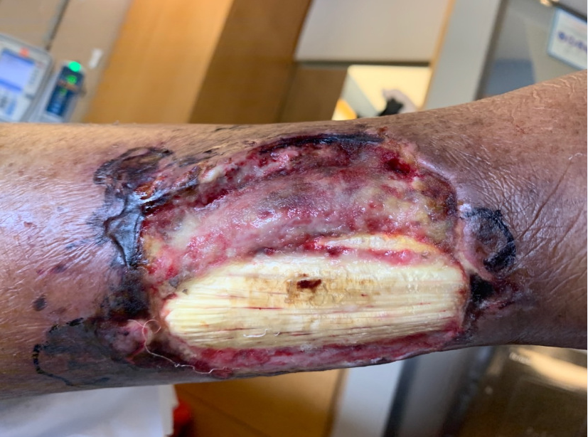
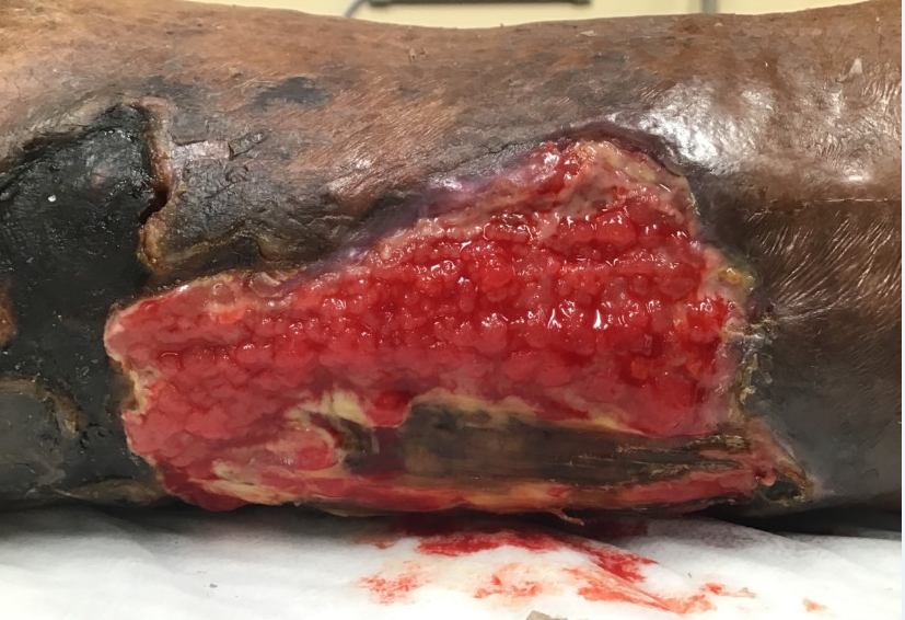
Case Presentation: An 80-year-old female with a history of atrial fibrillation on warfarin, type 2 diabetes mellitus, vitamin D deficiency, and CKD stage III presented as a transfer for further management of a worsening left lower extremity ulceration. Onset was 2 weeks prior without any concordant symptoms of infection and was initially evaluated by her PCP who had prescribed a course of cephalexin. Despite this, the ulceration had progressed with foul-smelling purulent drainage, swelling, and pain. At the outside institution CT of the left lower extremity was consistent with a superficial soft tissue swelling and calcifications with ultrasound negative for DVT. She was treated with broad spectrum antibiotics with negative infectious work up, and underwent wound debridement twice without clinical improvement. Patient was thereafter transferred to our institution with stable vitals. Labs illustrated a white blood cell count of 8.04 K/μL, Cr at 1.68 (at baseline), calcium of 9.0 mg/dL, PT/INR 1.8, CRP of 4.93 mg/dL, and ESR of 91 mm/hr. Warfarin was held on admission. Antibiotics were discontinued as there were no signs of cellulitis and repeat imaging did not reveal deeper infection. Dermatology was consulted and performed a shave biopsy illustrating calcific stippling of fat lobules and calcification of the walls of small and medium caliber vessels, consistent with calciphylaxis. With the diagnosis confirmed, vitamin D supplementation was discontinued and therapy with sodium thiosulfate was started. Pain control was achieved with low-dose opioids, wound care was provided by surgical teams, and anticoagulation was transitioned to apixaban. Fortunately, despite severe ulceration, there was no further progression during her hospital course. Patient was discharged to rehab and was noted to have significant improvement at follow up.
Discussion: Calciphylaxis is characterized by calcification of small and medium sized vessels that leads to ischemic necrosis (2). It classically occurs with renal failure but can also occur in those with normal renal function, precipitated by hyperparathyroidism, liver disease, corticosteroids, malignant neoplasm, or warfarin use (3). The exact mechanism is unclear but is thought to be promotion of vascular calcification by inhibition of vitamin K-dependent matrix Gla protein, a protein that prevents calcium deposition in arteries (4). Sodium thiosulfate is thought to have cation-chelating properties and therefore when used in calciphylaxis, chelating calcium to produce calcium thiosulfate which may be more soluble than other calcium salts (5).
Conclusions: Calciphylaxis is a rare condition associated with a mortality rate of up to 45% at 12 months from onset (1). It is prudent for clinicians to have a high index of suspicion, as it can also occur in non-end stage renal patients as highlighted by this case. Management requires an urgent multidisciplinary and individualized approach as there are no high quality studies supporting an optimal treatment regimen.