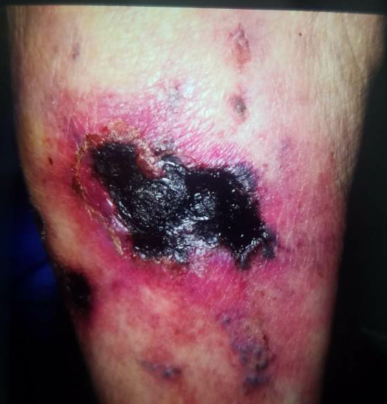
Case Presentation: 69 year old woman with atrial fibrillation on warfarin and unspecified connective tissue disease on prednisone presented to the emergency department (ED) with worsening, painful bilateral lower extremity skin lesions. She noted a mildly painful, non-healing left leg wound 3 months prior to presentation; then a 2nd similar lesion appeared on her right leg. She saw a physician who treated her with Bactrim and wound care for “chronic arterial/venous ulcer with superinfection” but her lesions enlarged and the pain worsened, leading to the ED visit. She denied fevers, chills, claudication, peripheral edema, and lower extremity trauma. Physical exam revealed a 7.4 cm left medial leg indurated eschar with serosanguinous drainage, and a 1.2 cm right posterior leg eschar without drainage. Laboratory data including serum creatinine, calcium-phosphate product, and parathyroid hormone levels were all normal. Inflammatory markers and uric acid levels were elevated. Left leg x-ray was unremarkable. Differential diagnosis included vasculitis, warfarin-induced skin necrosis, cold-precipitating proteins, stasis ulcer, severe peripheral arterial disease, neuropathic ulcer, and calciphylaxis. Skin biopsy of the right calf lesion was diagnostic of calciphylaxis. Warfarin was discontinued, prednisone tapered. She was started on pentoxyfylline, thrice weekly vitamin K and sodium thiosulfate, continuing at discharge with wound care and close dermatology follow-up.
Discussion: Calciphylaxis is a rare condition characterized by microvascular occlusions in adipose and dermal tissue, leading to painful, necrotic skin lesions due to tissue infarction. Typically seen in end-stage renal disease patients (“uremic calciphylaxis”), due to metabolic factors promoting extra-skeletal calcifications, but less commonly, it can be “non-uremic”. Primary hyperparathyroidism, malignancy, alcoholic liver disease, connective tissue disease, glucocorticoid and warfarin use are risk factors for non-uremic calciphylaxis, as are low serum albumin, elevated serum parathyroid hormone, phosphorus, and calcium levels. Our patient’s risk factors included glucocorticoid and warfarin use as well as her history of possible connective tissue disease. Her laboratory studies did not confer elevated risk. Non-uremic calciphylaxis is more commonly seen in females and has a predilection for the legs.
Conclusions: A high index of suspicion is required to diagnose non-uremic calciphylaxis as this condition is uncommon. Given the prevalence of atrial fibrillation and the resultant use of warfarin for anticoagulation, clinicians must be mindful of the fact that warfarin use is a key risk factor for this condition. Prognosis for non-uremic calciphylaxis is better than that of uremic calciphylaxis.