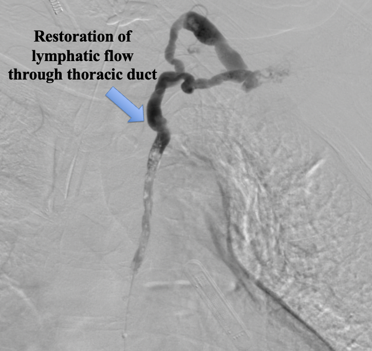
Case Presentation: A 54-year-old woman presented with six months of diarrhea and weight loss, and three months of abdominal distension. Her abdomen was distended and diffusely tender to palpation. Initial labs were notable for an albumin of 3.4 with normal bilirubin, AST, and ALT. Cross-sectional imaging demonstrated large ascites without evidence of liver pathology or ovarian mass. Her ascites was found to have a low SAAG with normal triglycerides and negative cytology x 2. Work up was otherwise notable for: LVEF 65% and no valvular disease on TTE, low-normal filling pressures on right heart catheterization, and UA negative for protein. Other pertinent labs included multiple nutritional deficiencies, elevated fecal fat, and positive spot stool alpha-1 antitryptase (A1AT). Negative work up included hepatitis B and celiac serologies, infectious stool studies, CEA/CA-125/CA 19-9, ANA, and peritoneal ADA. Given elevated fecal fat and stool A1AT, protein losing enteropathy (PLE) was considered. Upper/lower/capsule endoscopy revealed no identifiable cause of PLE. Interventional radiology was consulted for identification of possible chylous leak. MR lymphangiogram showed sluggish flow through thoracic duct (TD). TD angioplasty was performed, resulting in mildly improved TD flow post-procedure. Diarrhea resolved, and she was discharged with close outpatient follow up. She had recurrence of diarrhea and underwent repeat TD angioplasty with transient symptom resolution. When symptoms recurred again, she underwent thoracic duct bypass to external jugular (EJ) by plastic surgery with full symptom resolution and albumin normalization, and she has returned to her baseline weight.
Discussion: Ascites is a common presentation treated by hospitalists with a broad differential diagnosis, but our case was unusual in that it coincided with antecedent diarrhea and weight loss. The usual causes of ascites (CHF, cirrhosis, nephrotic syndrome) were quickly excluded in our patient, causing us to consider less common causes that would also explain her other symptoms. Our patient was found to have PLE, which prompted our consideration of an idiopathic TD obstruction.PLE is associated with primary and secondary intestinal lymphangiectasia that results in leakage of protein into stool. Primary etiologies include erosive and non-erosive gut pathology; secondary causes include lymphatic obstruction or conditions that increase lymphatic pressure (e.g., right heart failure). In this patient, the diagnosis of PLE was suspected given low albumin, diarrhea, and positive stool A1AT, in addition to unremarkable cardiac, hepatic, renal, and malignancy work ups. TD obstruction is a rare cause of PLE but should be considered when alternative causes have been excluded. The cause of the obstruction in our patient remains unclear.
Conclusions: Ascites requires a broad differential, with most common causes including hepatic, nephrotic, cardiac, and malignant etiologies. In cases where typical causes of ascites are ruled out, PLE with TD pathology is a reasonable diagnosis to consider. PLE requires extensive laboratory and endoscopic evaluation and, in the absence of underlying GI pathology, lymphangiography offers a specific tool for visualizing and excluding TD abnormalities, although it may not be readily available at most hospitals. Patients refractory to TD angioplasty may be candidates for thoracic duct bypass with IR and plastics.