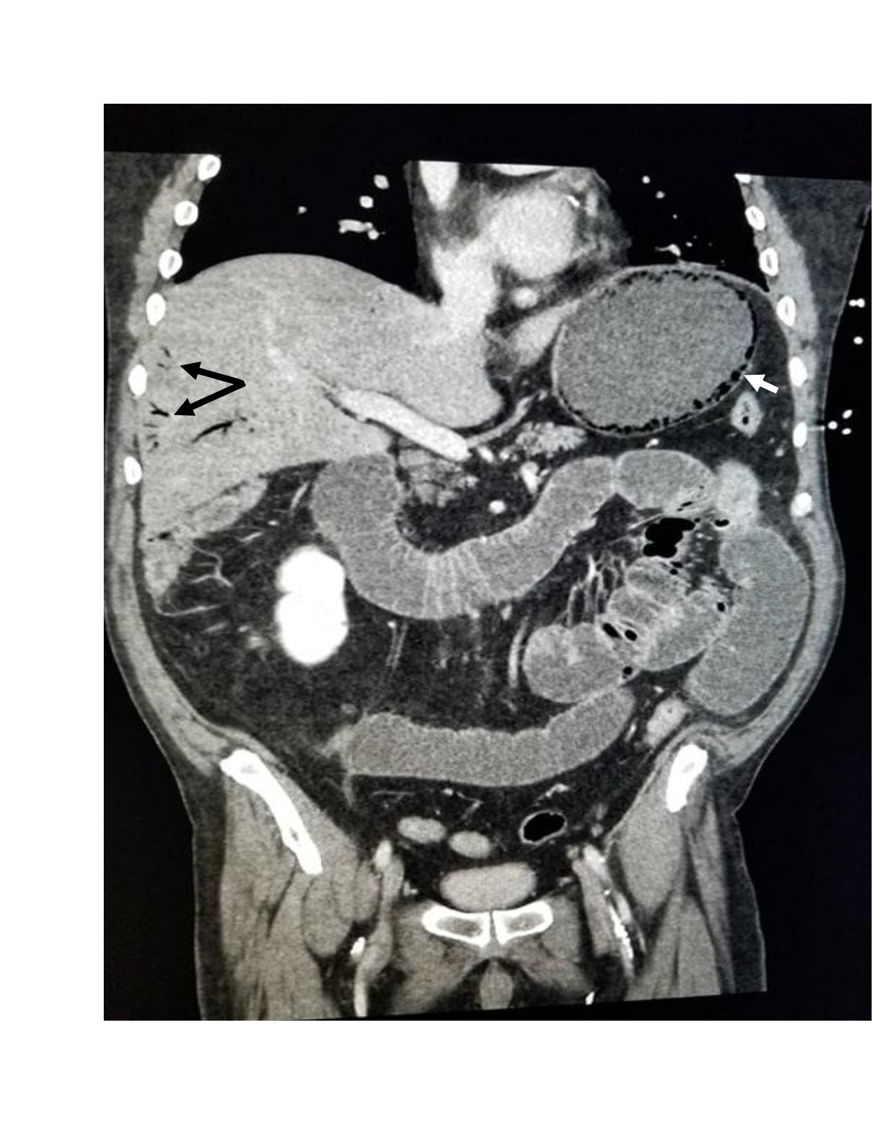
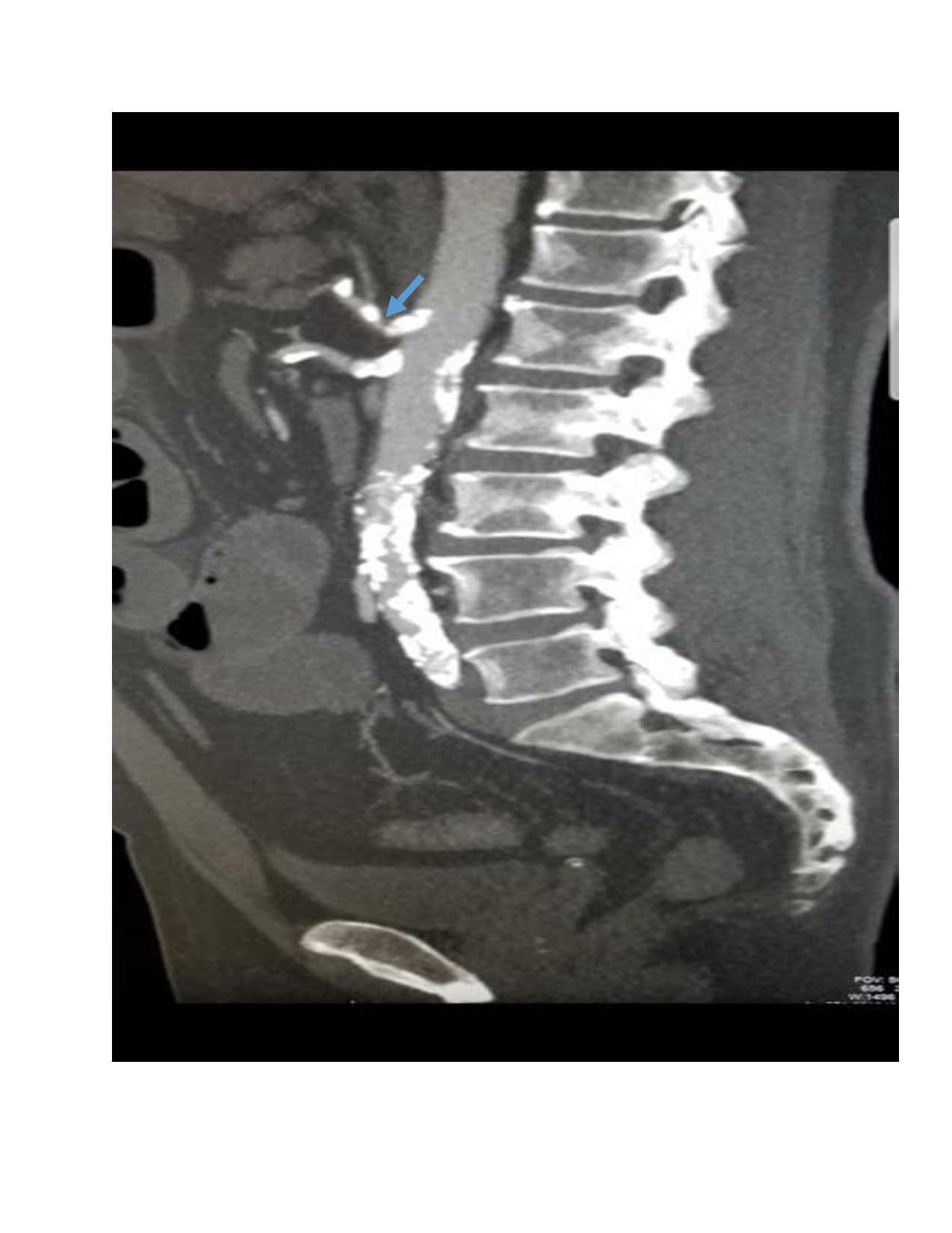
Case Presentation: A 64 year-old man with a history of coronary artery disease status post 5-vessel coronary artery bypass grafting and subsequent percutaneous coronary intervention, essential hypertension, hyperlipidemia, and type 2 diabetes mellitus (DM) presented to the hospital with a 4 month history of chronic progressive abdominal pain and diarrhea with a 70 pound unintentional weight loss. The patient had been admitted two weeks prior for the same complaints. A workup at that time included a computerized tomography (CT) scan of his abdomen and pelvis, colonoscopy and gastrointestinal (GI) PCR, which were all inconclusive. On this presentation, his pain has worsened and is predominantly mid-epigastric (MEG), left upper quadrant (LUQ) and left lower quadrant (LLQ). The pain is exacerbated by oral intake, improves slightly with a bowel movement (BM). It is associated with non-bloody diarrhea, his last BM being one day prior to admission, nausea and non-bilious, non-bloody vomitus. On exam, the patient was afebrile, heart rate 79 beats per minute, and blood pressure 159mmHg/68mmHg. His abdomen was diffusely tender, worse in MEG, LUQ and LLQ regions, with rebound, guarding and hypoactive bowel sounds. Laboratory results included a white blood cell (WBC) count of 21,000 per microliter with 7.3% bands and repeat negative GI PCR. CT angiography of the abdomen and pelvis revealed extensive atherosclerotic vascular disease with focal stenosis of the celiac, superior mesenteric artery (SMA), small bowel loop dilatation, and omental thickening. 24 hours later, repeat imaging showed emphysematous gastritis (EG) with gastric pneumatosis, venous gas surrounding the stomach and portal venous gas (Images). He was started on piperacillin-tazobactam and a nasogastric tube was placed. Vascular surgery placed a stent in the celiac and SMA and the patient had significant improvement in his pain. Antibiotics were deescalated to ceftriaxone and metronidazole. One week later, the patient was tolerating an oral diet and had return of his bowel function. He was discharged after completing 10 days of antibiotics.
Discussion: A rare but fatal variant of gastritis with a mortality rate as high as 60-80%, EG results from gastric invasion of gas forming organisms including Streptococci, Escherichia Coli, Enterobacter species, Clostridium Welchii, and Staphylococcus aureus. Factors causing disruption in the normal gastric integrity predispose to EG, including ingestion of corrosive substances, anti-inflammatory medications, DM, alcohol abuse, recent abdominal surgery, and ischemia. Radiologically, it can mimic gastric emphysema (GE), a non-infectious condition resulting from trauma or obstruction. Patients with GE and EG present with abdominal pain, nausea or vomiting. However, those with EG usually have severe abdominal pain, peritoneal signs and an elevated WBC count.We present a case of chronic progressive abdominal pain and weight loss consistent with chronic mesenteric ischemia, confirmed by CT angiography with celiac and SMA stenosis. Gastric infarction was subsequently complicated by infectious EG. The diagnosis of EG is radiographic and clinical with findings of gas in stomach wall and portal vein in a patient with severe abdominal pain and peritoneal signs.
Conclusions: Though emphysematous gastritis is uncommon, it is essential for hospitalists to recognize this rare form of gastritis in patients with severe abdominal pain in order to initiate early treatment given its high mortality rate.