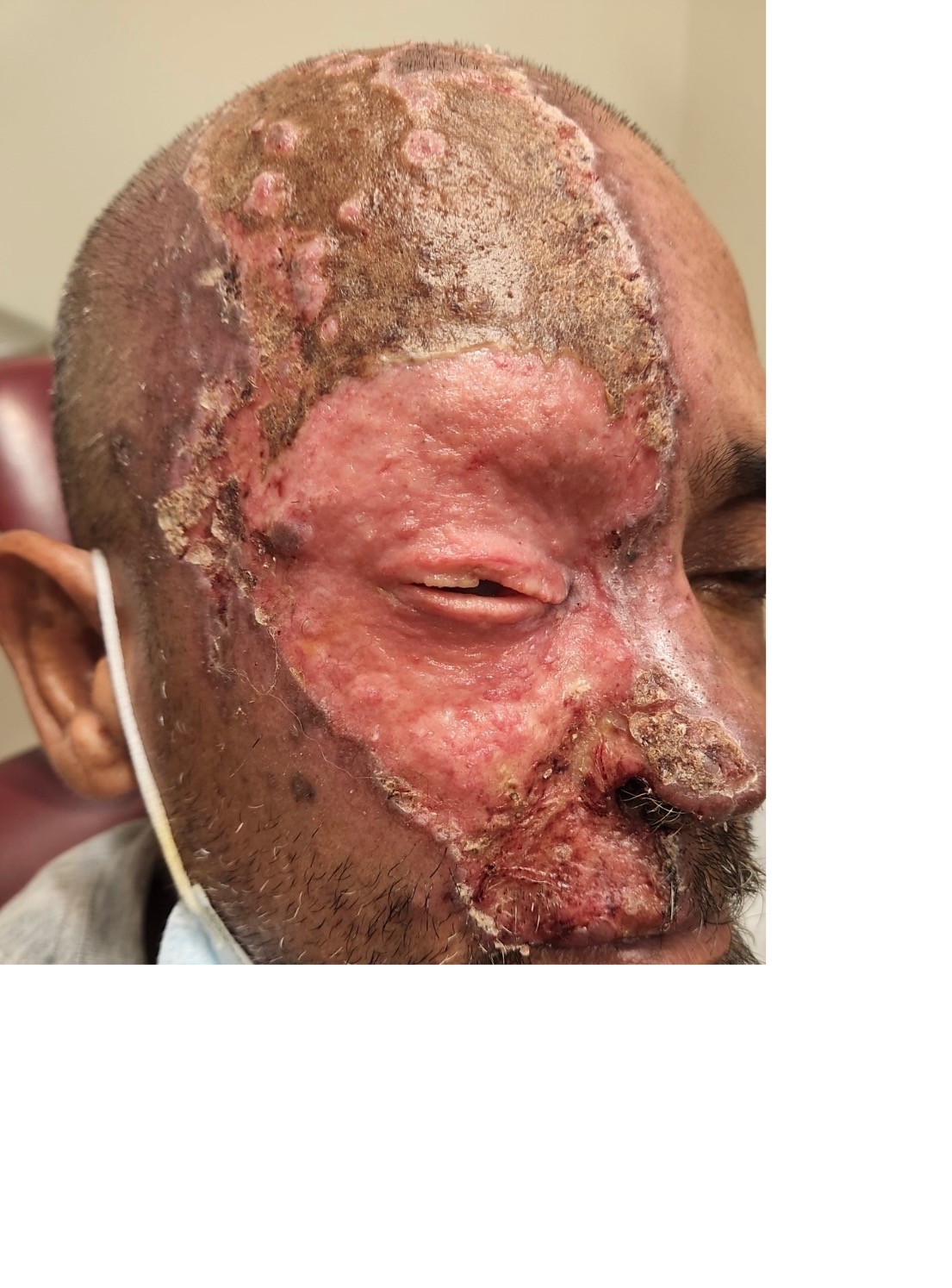
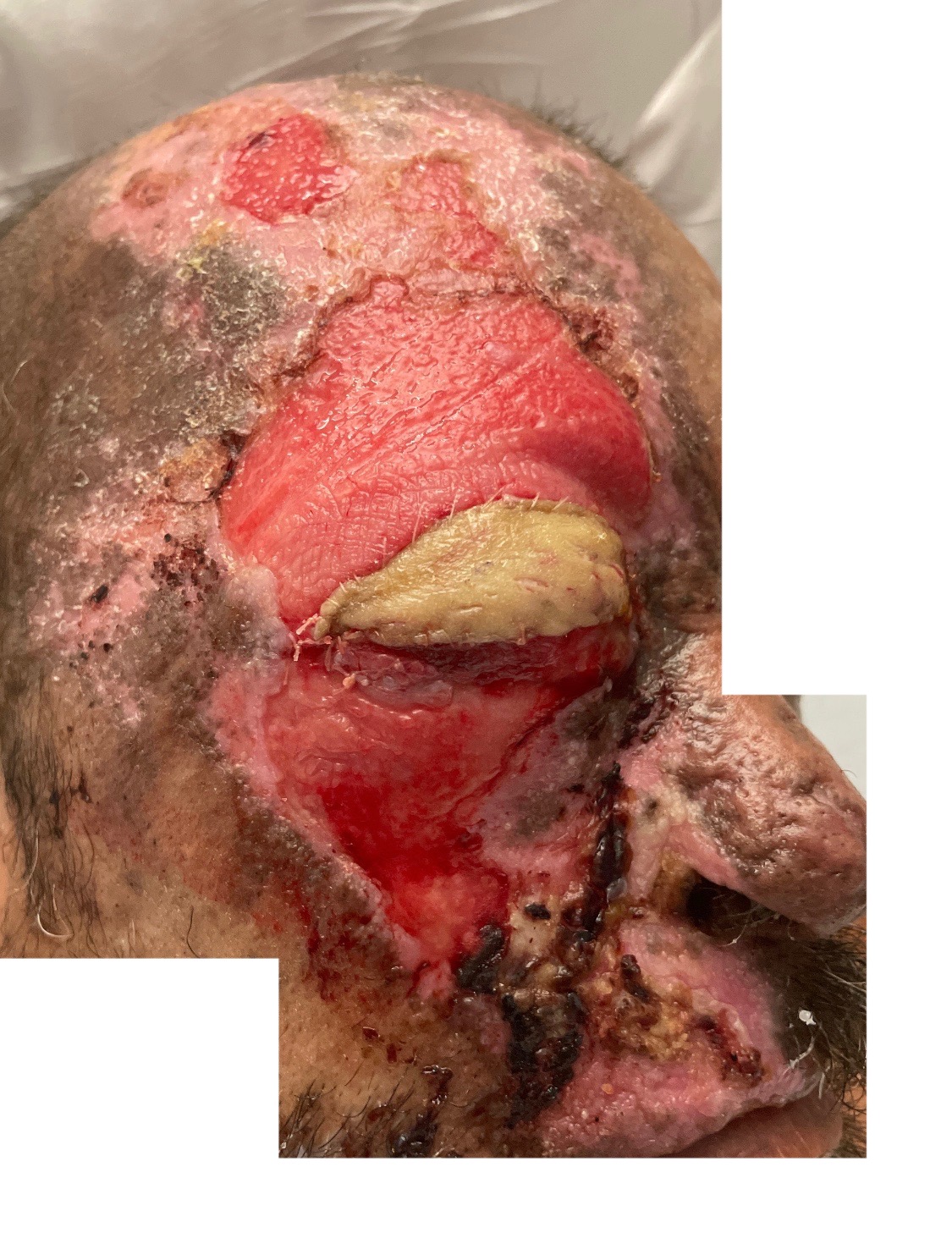
Case Presentation: A 58-year-old Spanish-speaking, uninsured man presents to primary care for a rash and is diagnosed with poison ivy and treated with prednisone. He eventually presents to the emergency department with ulcerative, crusting lesions of his right nasal ala, eyelid, scalp, philtrum, and lips. The lesions overlay a desquamative rash that covers his right face, extending from his scalp down to his upper lip, involving his right eyelid and eye including a corneal ulcer. Eye involvement also included hypopyon and necrotizing stromal keratitis. There is necrosis and destruction of his right nasal ala and destruction of his right upper and lower eyelids. Treatment was started with intravenous acyclovir, vancomycin and ampicillin-sulbactam and topical mupirocin and gentamicin. During his six day stay the patient was observed rubbing facial lesions despite counseling to stop. He was discharged on oral valacyclovir, cephalexin and gabapentin. He returned a week later for continued pain and worsening corneal ulcer. He was seen by ophthalmology, dermatology, psychiatry and the primary medicine team. After multidisciplinary discussion, ophthalmology performed an amniotic membrane graft with temporary tarsorrhaphy. The patient touched the ulcerated area, disrupting the temporary tarsorrhaphy. Ultimately, ophthalmology performed a permanent tarsorrhaphy. Upon extended counseling in Spanish by members of psychiatry and medicine, he adhered to further cares. He was discharged with gabapentin, oxycodone, duloxetine, doxycycline, gentamicin, Dermaphor ointment and dressings.
Discussion: Herpes zoster ophthalmicus is often seen in the elderly or the immunocompromised (1). Our case is distinct, occurring in an immunocompetent, 58-year-old. That his case was non-classic and he was not initially seen by an ophthalmologist or generalist with adequate expertise may have delayed his diagnosis and treatment. The severity of his ocular involvement is also rare. A review of 60 cases of trigeminal trophic syndrome (TTS) found 18% had corneal lesions and 16% had eyelid or canthal lesions (2). He not only had hypopyon, necrotizing stromal keratitis, and corneal ulcer requiring amniotic membrane graft, but also destruction of his upper and lower eyelids leading to permanent tarsorrhaphy with graft. His case was further complicated by poor communication due to language barriers compounded by inadequate pain and itching control leading to ongoing self-trauma. Of note, the language barrier was worsened by the patient’s fourth grade education level; initial clinicians were unaware of his limited education. Finally, unfamiliarity with TTS and lack of well-documented, effective treatments likely further exacerbated inadequate treatment and progression. Our case demonstrates a common infection progressing to a dramatic and rare outcome due to a combination of unlikely disease progression/presentation and several patient-specific barriers. When these factors are addressed, treatment success can be attained. After discharge, he returned to the emergency department for continued pain, however, the cutaneous lesions and graft were stable and healing. He follows with ophthalmology.
Conclusions: Herpes zoster ophthalmicus is a rare manifestation of herpes zoster that should be considered in immunocompetent individuals. Language, insurance, and educational barriers add significant difficulty to successful treatment, but prompt analgesia and patient-specific education is essential to management.