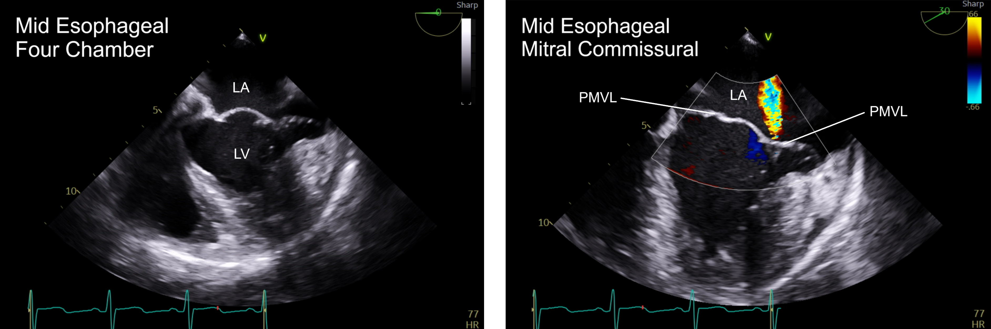
Case Presentation: A 52-year-old man with no known medical history presented to the emergency department with four days of worsening neck pain, subjective fevers, fatigue, and new-onset palpitations. He met SIRS criteria for sepsis and was started on empiric broad-spectrum antibiotics with concern for meningitis. CSF studies and cervical spine MRI were unremarkable. During hospitalization, he was intermittently tachycardic but otherwise hemodynamically stable. An EKG revealed atrial flutter with 2:1 conduction, and transthoracic echocardiogram revealed moderate-to-severe mitral regurgitation, mitral prolapse, and a markedly dilated left atrium. His differential included viral myocarditis, a post-viral syndrome, and occult tick-borne illness; however, viral studies all returned negative. Despite supportive care and rate control with diltiazem, the etiology of the patient’s atrial flutter and systemic symptoms remained unclear. After several days, a senior clinician noted at the bedside the patient’s striking height and slender habitus which prompted a radical revaluation of his clinical findings. The patient was 6’7 with pronounced hypermobility of multiple joints including positive wrist and hand signs bilaterally, suggestive of a possible connective tissue disorder, most likely Marfan Syndrome. The patient’s symptoms improved with diltiazem and on follow-up 1 month after discharge, he reported complete resolution of his palpitations and neck stiffness. Arrangements were made for formal genetic testing.
Discussion: Marfan syndrome is an autosomal dominant connective tissue disorder caused by mutations in fibrillin-1, a glycoprotein essential for extracellular matrix integrity and TGF-β signaling. It commonly involves the cardiovascular, skeletal, and ocular systems, with cardiovascular complications such as mitral valve prolapse and aortic aneurysm being most life-threatening. Atrial arrhythmias occur in ~16% of patients.While our patient’s neck pain and atrial arrhythmia were initially concerning for sepsis due to suspected meningitis, no infectious source was ever identified. In retrospect, Marfan syndrome could plausibly explain several elements of this patient’s presentation, including his dilated left atrium, mitral valve prolapse, and rapid-onset atrial flutter. His neck pain may have also represented cervical ligamentous laxity or postural strain, particularly in the cervical spine where spondylosis was noted. Importantly, his skeletal findings of positive wrist/thumb signs, mitral valve prolapse, and high palate yield a Ghent score 5 (moderate risk). While he lacked family history or known ectopia lentis, he met criteria for genetic testing.
Conclusions: This case highlights the enduring importance of bedside examination. In an era of increasing reliance on imaging, laboratory data, algorithms, and artificial intelligence, the physical exam remains essential for shaping clinical reasoning. In this case, the diagnosis of Marfan syndrome may have been delayed or missed entirely had the team relied solely on imaging and laboratory results. The recognition of Marfan syndrome reframed the clinical picture and led to appropriate outpatient referrals for genetic and cardiac evaluation.