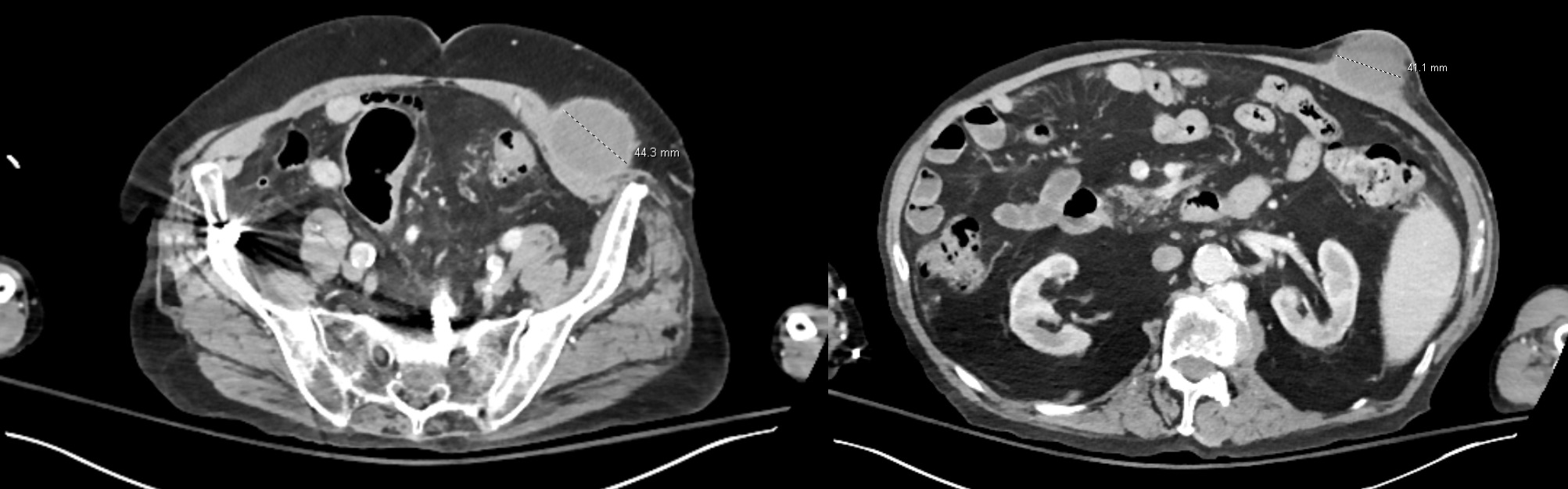
Case Presentation: Nocardiosis is an opportunistic infection caused by aerobic, weakly acid-fast, branching gram-positive organisms of the genus Nocardia. The disease typically begins as a pulmonary infection and disseminates hematogenously to the central nervous system (CNS) and other organs.¹ Nocardia farcinica is particularly virulent, with higher rates of dissemination and CNS involvement.² Primary cutaneous nocardiosis originating from the abdominal wall and progressing to disseminated disease is extremely rare.³ Early recognition and prompt CNS imaging are essential to reduce morbidity.¹ We present a rare case of disseminated N. farcinica initially manifesting as an abdominal wall abscess in an immunocompromised patient.An 82-year-old male with sarcoidosis on chronic corticosteroids, chronic kidney disease 3b, and a remote neurosurgical history presented with two weeks of progressive left upper-quadrant abdominal wall swelling and pain unresponsive to oral antibiotics. He was lethargic but hemodynamically stable. Labs showed WBC 12.2×10⁹/L, hemoglobin 11.8 g/dL, platelets 108×10⁹/L, and creatinine 2.2 mg/dL. CT abdomen revealed a 4.1-cm left rectus muscle abscess and a 4.4-cm rim-enhancing abscess within the left pelvic abdominal wall musculature. The rectus abscess was drained at bedside, and the patient underwent operative incision and drainage for the pelvic abscess with collection of purulent fluid was obtained from both. Empiric vancomycin and piperacillin-tazobactam were started. On hospital day 4, cultures from both abscesses grew Nocardia farcinica. The patient also reported two months of progressive confusion, so MRI brain was obtained which demonstrated multiple bilateral frontal lobe ring-enhancing lesions with restricted diffusion and vasogenic edema, consistent with intracranial abscesses. CT chest revealed a 2.3×0.9-cm right lower-lobe pulmonary nodule with internal lucency, concerning for pulmonary nocardiosis. He was transitioned to high-dose IV trimethoprim-sulfamethoxazole and imipenem-cilastatin. Neurosurgery recommended transfer to a tertiary center. The patient remains hospitalized, clinically improving, and undergoing repeat neuroimaging.
Discussion: This case highlights disseminated N. farcinica with an atypical initial presentation as an abdominal wall abscess—an uncommon manifestation reported only rarely.³ Chronic corticosteroid use was the major predisposing factor. The case underscores the importance of considering nocardiosis in immunocompromised hosts with deep soft-tissue infections unresponsive to standard antibiotics.⁴ Because N. farcinica shows neurotropism and is more likely to cause CNS abscesses, brain MRI is essential in all confirmed infections, even with minimal neurologic symptoms.³ Prolonged dual antimicrobial therapy with CNS-penetrating agents is required, and early diagnosis with multidisciplinary management is crucial to reduce morbidity and mortality.⁵
Conclusions: Disseminated nocardiosis may rarely initially present with soft-tissue abscesses. Early CNS imaging and prompt initiation of dual therapy are critical for reducing morbidity in immunocompromised patients with suspected Nocardia infection.