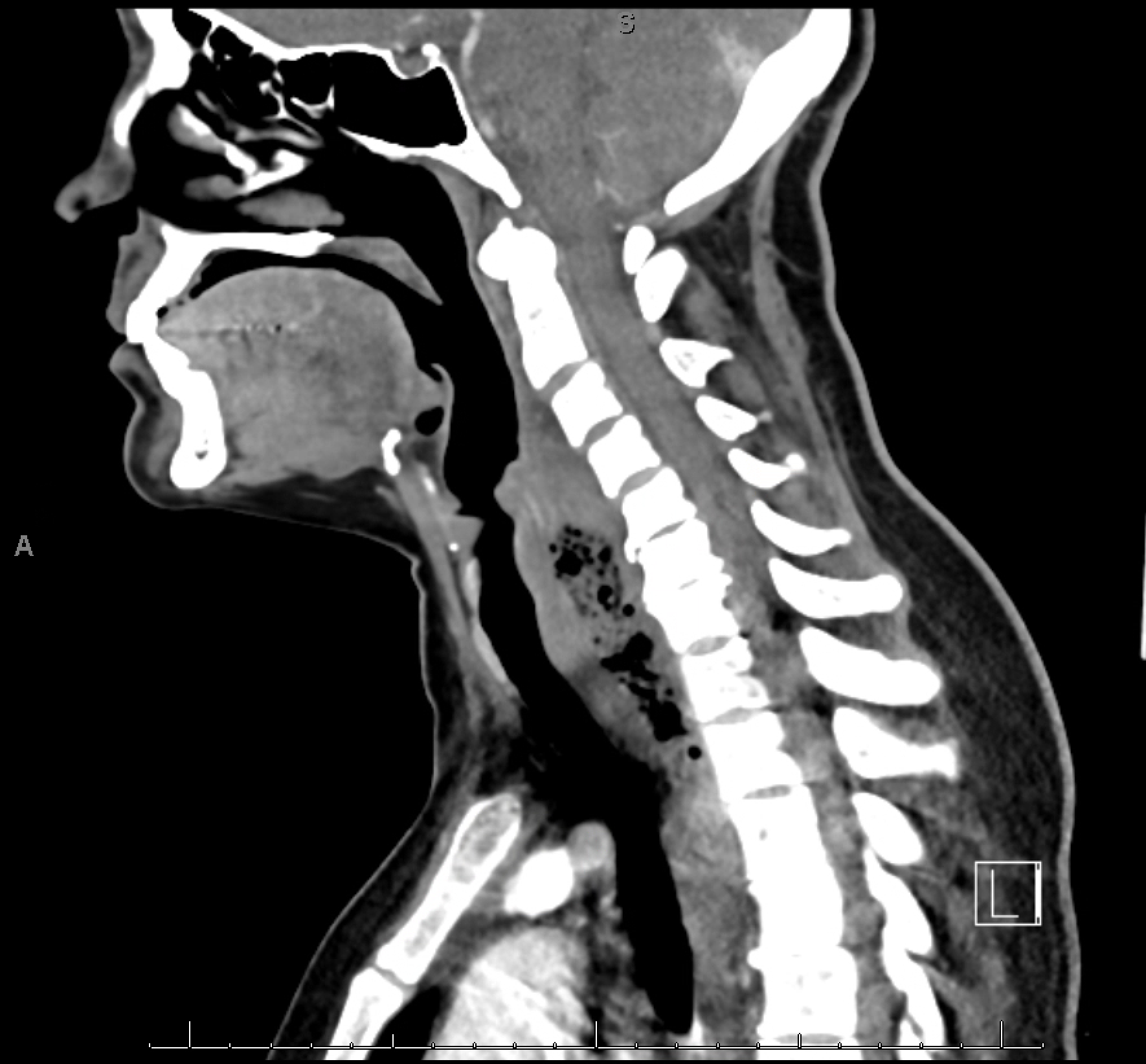
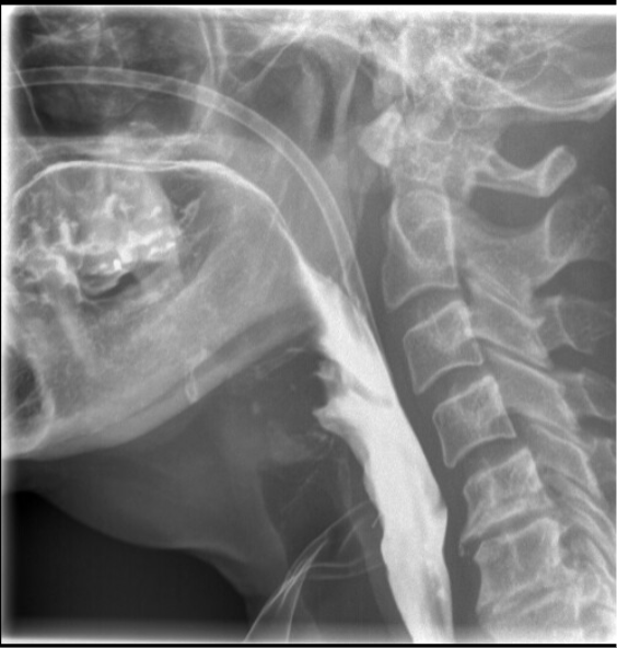
Case Presentation: A 50-year-old female with May-Thurner Syndrome presents with three days of odynophagia following consensual “aggressive” oral sex. Barium swallow study confirmed cervical esophageal perforation; CT neck & chest found mediastinal free air. She underwent incision & drainage with pharyngoesophageal repair with Otolaryngology (ENT) and received prolonged IV antimicrobials. Course was complicated by an esophageal leak and drain dislodgement requiring a second ENT intervention. POD 25 esophagram demonstrated no leak; thereafter she resumed oral intake. This is the first case of esophageal perforation following consensual oral sex and highlights the importance of early recognition and intervention for favorable patient outcomes.
Discussion: Oral intercourse as a cause of esophageal perforation is an exceptionally rare presentation with this being the first documented case of perforation following consensual oral sex, other cases in the literature are unfortunately the result of nonconsensual oral sex. Most cases secondary to this mechanism involve a hypopharyngeal injury or a proximal esophageal rupture and the presentation is usually immediately after the trauma. Our patient presented three days after her injury with a posterior cervical perforation (C5-C6) and required staged surgical interventions due to ongoing contamination and poor tissue quality. Intraoperative findings revealed a tear through the mucosa but contained within the muscular layer. The mucosal tear allowed for collection of food and saliva between the mucosa and muscularis layers of the esophagus requiring drainage intraoperatively. The likelihood of repair on the first attempt would have been higher had the patient presented immediately following the injury as the mucosal layer would have had more structural integrity. Continuing oral intake likely enlarged the mucosal defect which further compromised the repair integrity. For this case, the weak, poorly vascularized mucosal layer led to reaccumulation of substance between the esophageal layers requiring a second operation for washout and repair. From a diagnostic perspective, NPL cannot reliably visualize beyond the hypopharynx making it not the ideal form of evaluation for this patient resulting in disposition to the internal medicine team when she would have been better served as an otolaryngology primary patient.
Conclusions: The mechanism of injury is important and seemingly low-energy etiologies (e.g., oral intercourse) can still cause significant esophageal injury. In this case, further history clarified the potential energy of the trauma as the true mechanism of action was likely different than assumed by clinicians at urgent care, OSH ED, our ED, and our admitting team which may have conferred different pre-test probability and, thus, initial urgency of diagnostic and management. Further, contained perforations can be more challenging to repair given the integrity of the mucosa with contamination and collection between esophageal layers. Delayed presentation causes tissue quality decline, more difficult repair, and possibly staged interventions leading to a worsened prognosis. Early surgical involvement is critical, and specialty is usually determined by location of esophageal injury. This case reinforces that early recognition, anatomical localization, and multidisciplinary coordination are essential to achieving favorable outcomes in patients with an esophageal perforation.