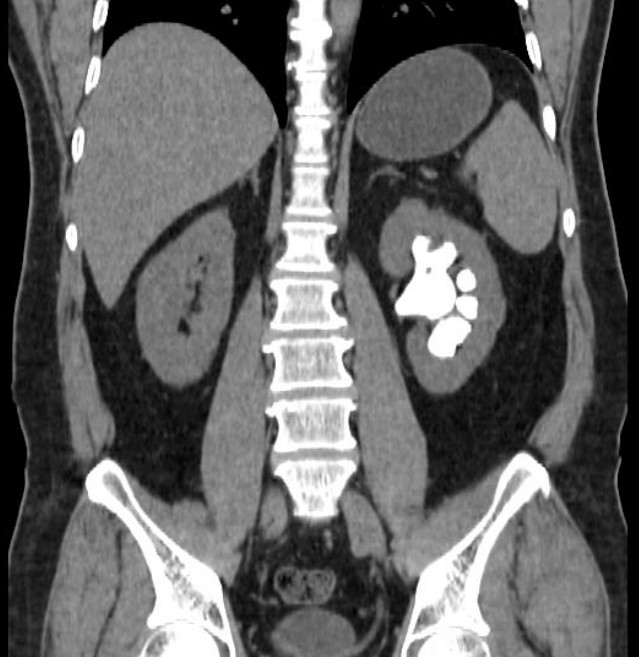
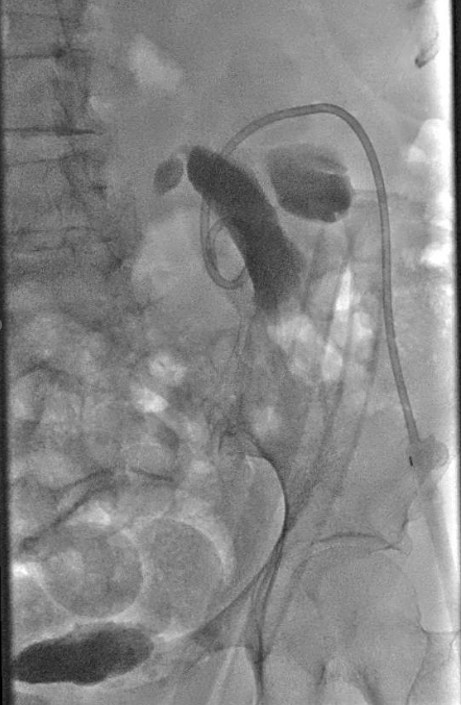
Case Presentation: A 67yo woman with severe COPD paroxysmal AFib presented with acute chest pain and dyspnea, leukocytosis and acute-on-chronic hypoxemic respiratory failure. A CT angiography of the chest showed a left upper lobe infiltrate and no pulmonary embolism. She was admitted and treated for COPD exacerbation secondary to pneumonia.On hospital day 2, she developed flank pain. A non-contrast CT abdomen showed a large branching hyperdense structure occupying the right renal pelvis and extending into multiple calyces, associated with moderate hydronephrosis, and interpreted as a staghorn calculus (fig 1). The patient had no history of nephrolithiasis, recurrent urinary tract infections, dysuria, or hematuria, but did report intermittent right lower quadrant pain over the past year. She also reported a prior history of “ureteral narrowing” . Nuclear renography showed asymmetric renal function, with 29% uptake in the right kidney and 71% in the left. Urology and IR services recommended staged right percutaneous nephrolithotomy.Intraoperatively, only a small renal pelvic stone was identified and removed, and a significantly chronically dilated right renal pelvis with no evidence of staghorn calculi was noted. An antegrade nephrostogram (fig 2) confirmed ureteral patency without filling defects.Careful review of prior imaging demonstrated a high-inserting ureteropelvic junction with chronic pelvic dilation. The apparent “staghorn calculus” corresponded to retained iodinated contrast from the CTA performed 48h earlier. An antegrade nephrostogram confirmed ureteral patency without filling defects. A nephroureteral stent was placed, and the patient subsequently underwent right nephrostomy tube placement for persistent hydronephrosis. She improved clinically and was discharged with plans for outpatient evaluation for possible pyeloplasty.
Discussion: This case highlights an uncommon but important pitfall in renal imaging: retained contrast mimicking staghorn calculi. On non-contrast CT, hyperdense retained contrast within a poorly draining collecting system may appear indistinguishable from stone material, particularly when it branches through the calyces in a pattern typical of staghorn calculi. Patients with abnormal renal anatomy, such as ureteropelvic junction (UPJ) obstruction, are especially prone to this artifact due to contrast pooling. Careful review of recent imaging, attention to attenuation values, and awareness of timing relative to contrast-enhanced studies are critical to avoiding misdiagnosis. In this case, the mimicry led to more aggressive surgical intervention before intraoperative findings revealed the true etiology. Greater vigilance among radiologists, urologists, and hospitalists can help prevent such misdiagnoses and unnecessary procedures.
Conclusions: Retained contrast in a dilated collecting system can mimic staghorn calculi on non-contrast CT, leading to potential diagnostic and therapeutic errors. Coordination and communication among radiology, urology, and medicine teams improves diagnostic accuracy and patient outcomes.