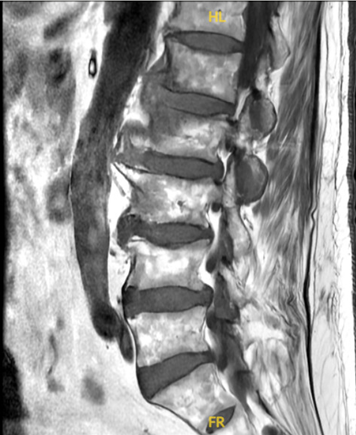
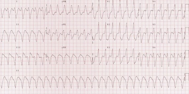
Case Presentation: A 79-year-old Caucasian male presented with a two-week history of progressively worsening low back pain. The pain was unresponsive to rest or over-the-counter analgesics and had become severe enough to limit ambulation. He denied any history of trauma, fever, or recent infections.His past medical history was significant for hypertension, coronary artery disease, hypothyroidism, peripheral arterial disease, prostate cancer (status post-prostatectomy), stage 3 chronic kidney disease, osteoarthritis, and sick sinus syndrome status post pacemaker placement.On physical examination, the patient was afebrile and hemodynamically stable. Neurological examination revealed no focal deficits, but straight leg raise was positive bilaterally. Musculoskeletal exam noted midline tenderness from L1 to L5 without signs of epidural compression. There were no skin lesions, rashes, or other evidence of systemic infection.Laboratory studies showed a white blood cell count of 16.0 ×10⁹/L. Inflammatory markers were within normal limits (ESR 18 mm/hr; CRP 2.7 mg/L). Blood cultures drawn on admission returned positive for Enterococcus faecalis in two separate sets. Due to high clinical suspicion, a CT-guided biopsy of the lumbar spine was performed, which confirmed E. faecalis infection.MRI of the lumbar spine demonstrated multilevel spondylosis and canal narrowing at L4–L5. There was only subtle T2 prolongation and mild enhancement at the L1 and L2 vertebral endplates—findings that were initially considered nonspecific.The patient was started empirically on IV vancomycin and ceftriaxone. Following culture results and antibiotic susceptibility testing, E. faecalis was found to be susceptible only to ampicillin. Despite a remote history of penicillin allergy, a monitored challenge was successfully conducted, and ampicillin was initiated.Given the presence of an indwelling pacemaker, a transthoracic echocardiogram was performed, which was negative for vegetations. However, during preparation for a transesophageal echocardiogram (TEE), the patient developed ventricular tachycardia and was transferred to the cardiac ICU. A pacemaker infection was suspected; the device was removed and replaced with a temporary pacing system during antibiotic therapy. After stabilization, a new permanent pacemaker was implanted.The patient’s back pain and weakness improved notably with targeted antibiotic therapy. Repeat imaging showed no new findings, and he was discharged in stable condition to a rehabilitation facility.
Discussion: This case demonstrates E. faecalis-induced vertebral osteomyelitis with initially negative MRI. Although MRI sensitivity is high (~90%), false negatives occur, especially early in infection or with low-virulence organisms. Subtle imaging changes may be missed or develop later.Clinical suspicion remains paramount despite negative imaging. Elevated WBC and positive cultures confirmed diagnosis. Hardware infections, such as pacemaker-associated infections, require prompt diagnosis and management, as Enterococcus readily colonizes foreign bodies.
Conclusions: Spinal osteomyelitis should be considered in elderly patients with persistent back pain and elevated inflammatory markers, even if imaging is inconclusive. Enterococcus species, though rare, are important pathogens. Early biopsy, culture, and targeted antibiotics are essential. Source control, especially with implanted hardware, is critical to prevent persistent or recurrent infection.