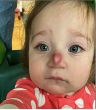
Case Presentation: A healthy 15‑month‑old female with a congenital nasal lesion presented to the Emergency Department with one week of progressive swelling and erythema of the lesion. She was afebrile, with one day of congestion and no other systemic symptoms. Oral cephalexin prescribed two days earlier had no effect. There was no history of trauma, drainage, or prior skin infections. The lesion, present since birth, had been diagnosed as a juvenile xanthogranuloma in the outpatient setting, with plastic surgery follow‑up and plans for excision after age three. Vital signs were appropriate for age. On examination, the lesion was erythematous without drainage or an external sinus tract; the remainder of the exam was unremarkable. Laboratory studies – including complete blood count, basic metabolic panel, and C‑reactive protein – were normal. She tested positive for rhino/enterovirus and negative for MRSA. Intravenous ampicillin/sulbactam was initiated, and plastic surgery was consulted. The differential diagnosis included juvenile xanthogranuloma, nasal dermoid cyst with cellulitis, and dermoid cyst with sinus tract. Swelling improved with antibiotics, though erythema persisted. MRI and CT of the head revealed a midline nasal mass with a sinus tract extending to the foramen cecum. The patient remained neurologically stable and was discharged on hospital day 7 with oral amoxicillin/clavulanic acid. Two weeks later, neurosurgery and plastic surgery performed a successful excision of the nasal dermoid with intracranial sinus tract extension. She was discharged on a 7‑day course of cephalexin and remained clinically well.
Discussion: Nasal dermoid cysts with sinus tracts are rare congenital anomalies arising from ectodermal inclusion during embryogenesis. These lesions often contain skin appendages and typically present as midline nasal masses in early childhood, though some remain undetected until later in life. While many are confined superficially to the nasal bones, up to 45% extend intracranially through sinus tracts, placing patients at risk for recurrent infection, drainage, or intracranial complications. Accurate diagnosis is critical because management hinges on defining the full extent of the lesion. MRI and CT are essential to delineate intracranial involvement and guide surgical planning. Inadequate evaluation may lead to incomplete excision, recurrence, or missed intracranial extension with serious consequences. In this case, the presence of overlying cellulitis prompted early imaging, which revealed a sinus tract that would otherwise have remained undetected. This underscores how clinical progression can serve as a trigger for timely imaging and appropriate multidisciplinary intervention.
Conclusions: Midline nasal lesions require thorough evaluation to distinguish dermoid cysts and identify potential sinus tracts. Because treatment decisions – including the need for advanced imaging and complete surgical excision – depend directly on diagnostic accuracy, early recognition is essential. Imaging should be pursued promptly in the setting of infection, atypical progression, or diagnostic uncertainty. Multidisciplinary management ensures safe and definitive resection, preventing recurrence and avoiding potentially severe complications from missed intracranial extension.