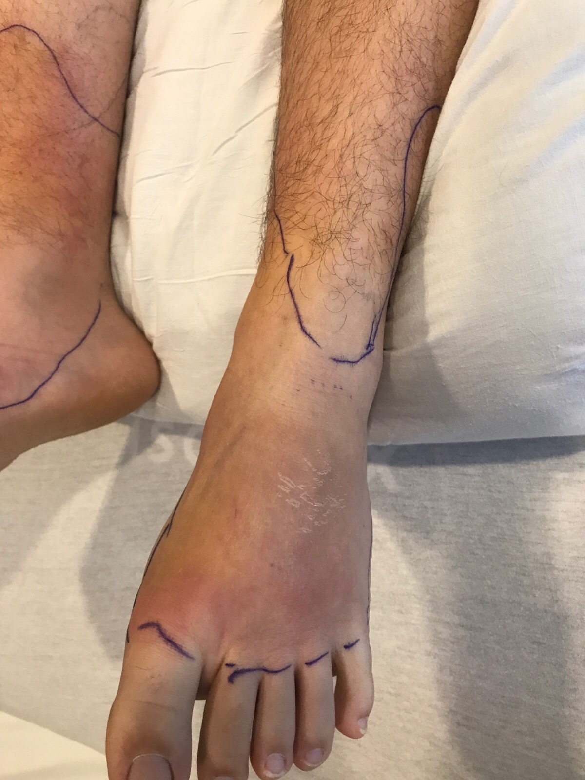
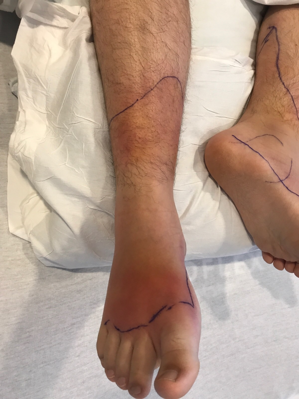
Case Presentation: A 17 year-old male teenager presented with two weeks of bilateral lower extremity edema, erythema, and warmth associated with fevers and joint pain at the foot and ankle. After a presumed bug bite on his left foot he developed a rash that spread from left to right leg. Symptoms did not improve with Doxycycline. Patient was hospitalized and treated for bilateral lower extremity cellulitis with IV Vancomycin and Clindamycin. On admission, he was febrile and had an erythematous confluent rash with swelling at bilateral feet extending up to past the ankles. There was exquisite pain with walking. MRI and ultrasound were negative for osteomyelitis and deep-vein thrombosis. He was discharged on PO Clindamycin but continued to have fevers. He was re-admitted and did not have clinical improvement on Vancomycin. Pediatric Infectious Disease, Dermatology, and Rheumatology were consulted due to suspicion for inflammatory or autoimmune process. Skin biopsy revealed septal panniculitis consistent with erythema nodosum. Chest X-ray and Chest CT demonstrate bilateral hilar lymphadenopathy, which was later biopsied and was positive for non-caseating granuloma consistent with sarcoidosis.
Discussion: Cellulitis is incorrectly diagnosed in as many as 1/3 of patients. One study estimates cellulitis misdiagnosis leads to 50,000 to 130,000 unnecessary hospitalizations and unnecessary antibiotics causing roughly 9,000 nosocomial infections annually.
Diseases that commonly masquerade as infectious cellulitis include stasis dermatitis, lymphedema, lipodermatosclerosis, irritant dermatitis, superficial thrombophlebitis, deep vein thrombosis, eosinophilic cellulitis, gout, Sweet syndrome, sarcoidosis, and panniculitis.
In the absence of trauma to both legs, bilateral cellulitis is exceedingly rare. Lower extremity cellulitis is caused by direct inoculum to an affected limb. Bilateral cellulitis would require either bacterial dispersion or independent inoculum of both legs. Thus the diagnosis of “bilateral cellulitis” should prompt clinician to look for noninfectious causes. A skin biopsy may be more helpful early in the course before advanced diagnostic tests are considered.
The literature lacks data about the relative frequency of infectious cellulitis compared with non-infectious mimickers of cellulitis. ALT-70 is a model predictive of lower extremity cellulitis recently developed to facilitate more accurate diagnosis: Asymmetry (3 points), Leukocytosis (1 point), Tachycardia (1 point), and age ≥70 (2 points). Score of 0-2 points indicate ≥83.3% likelihood of pseudocellulitis and ≥5 points indicate ≥82.2% likelihood of true cellulitis.
Conclusions: Cellulitis is a common misdiagnosis leading to unnecessary antibiotics use and hospitalization. It is important to keep a broad differential and to have high clinical suspicion for noninfectious causes of lower extremity erythema as to not make the mistake of diagnosing “bilateral cellulitis”.