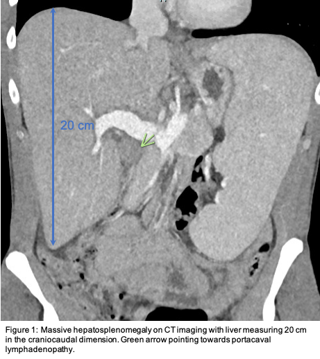
Case Presentation: 37 year-old male with a medical history significant for intravenous drug usage presented with three years of progressive generalized symptoms including weight loss, fevers, and shortness of breath. The patient started to experience worsening fatigue, drenching night sweats and an unintentional weight loss exceeding 20 pounds over several months leading to admission. Upon initial workup patient was noted to have elevated alkaline phosphatase of 893 (40-150 U/L), aspartate aminotransferase 277 (5-35 U/L) and alanine aminotransferase 154 (0-55 U/L), pancytopenia with white blood cells of 2.3 x 10³ (3.5-10.5×10³/uL), hemoglobin 11.8 (13.5-17.5 g/dL) and platelets 108 (150-400 x 103/uL). This prompted imaging with computerized tomography of the chest, abdomen and pelvis (Figure 1) revealing massive hepatosplenomegaly with portacaval lymphadenopathy and periportal edema along with multiple bilateral peripherally based pulmonary nodules largest being 5 mm. An extensive workup including autoimmune, viral, mycobacterial serologies and screening oncologic labs resulted within normal limits to which a liver biopsy was obtained depicting non-caseating granulomas. During the work up a (1,3)-beta-D-glucan resulted >500 (Negative: <60 pg/mL) along with Coccidioides Antibody IgG 1.4 (Negative: <0.9 IV). These findings with an uncovered history of frequent travel to the southwest, allowed for the diagnosis of disseminated coccidioidomycosis to be established.
Discussion: Coccidioidomycosis is caused by the fungi Coccidioides immitis and posadasii which are classically known for its geographic prevalence in the deserts of the southwestern United States, where spores produced by Coccidioides remaining in the air for prolonged periods, can lead to pulmonary penetration and disease. Although a large majority of patients are asymptomatic, many present with generalized cough, shortness of breath and fevers with a rare number escalating to drenching night sweats and weight loss once progressed to extrapulmonary infection via hematogenous spread. Disseminated coccidioidomycosis is rare in the general population, <1%, with an increased risk for immunocompromised hosts; interestingly this case presents an immunocompetent patient. Common sites of infection include soft tissue (cutaneous granulomatous lesions), bone (vertebral lesions), central nervous system (meningitis) and as in this case the hepatosplenic system. Treatment includes Itraconazole or Fluconazole for a minimum 3 to 6 months with severe cases (disease of cranium and vertebral bodies) being targeted with Amphotericin B.
Conclusions: Disseminated coccidioidomycosis is a rare disease process that can linger undetected for months to years before ‘B symptoms’ and affected tissue changes lead to hospitalization, which can be difficult to diagnose and important to keep in the differential. This case presents a very rare form where the granulomatous process led to massive hepatosplenomegaly and extensive workup prior to (1,3) – beta-D-glucan unmasking the etiology.