Case Presentation: 76 year old female with a history of COPD, Asthma, Allergic Rhinitis, and GERD presented to the Emergency Department for evaluation of worsening fatigue, dysgeusia and weight loss. Given these symptoms, the patient’s allergist referred her for COVID-19 testing. The patient denied any respiratory or GI symptoms, and review of symptoms was only notable for a left sided chest/flank pain. She was sheltering in place and denied sick exposure or travel. She was a former smoker with a 40 pack year history. PAP smear, mammogram and colonoscopy were up-to-date and normal. On presentation, vital signs were notable for a low-grade fever of 100.7 F and tachycardia to the 120s. Oxygen saturation was 99% on room air. Her admission labs were remarkable for a hemoglobin of 7.8 g/dL, down from a baseline of 12.6 g/dL six months prior and a mild thrombocytopenia of 107,000. SARS2 COVID nasal PCR was negative. She had a mild elevation of inflammatory markers: CRP 12.6 ng/dL and ESR 33 mm/hr, Interleukin-6 was 37.94 pg/mL. She also had a mild coagulopathy with INR 1.35, PT 15.4s and PTT 32s. D dimer was elevated at 3028 mg/dL. Chest X-Ray showed scattered, ill-defined nodular airspace opacities. Cardiac workup with EKG and troponins were negative for ischemia. A CT Angiogram of the chest ruled out pulmonary embolus but again suggested a multifocal pneumonia .
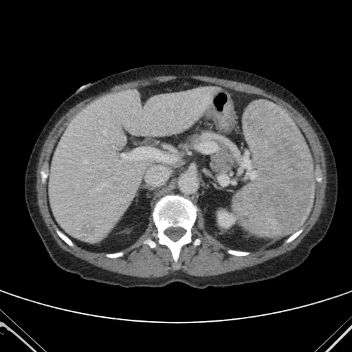
Discussion: Patient had a comprehensive inpatient workup. Admission blood cultures and sputum cultures were no growth. Testing for Legionella pneumophilia, Streptococcus pneumoniae and common viral pathogens were negative. A repeat COVID-19 test was negative. Hospital course was complicated by acute anemia requiring multiple transfusions. Hematologic workup was negative for overt bleeding, hemolysis, B12/folate deficiencies or hematologic malignancy. A CT abdomen/pelvis showed marked splenomegaly with heterogeneous enhancement and enlarged mesenteric lymph nodes. Given the bicytopenia and splenomegaly, HIV, HAV, HBV, HCV, CMV, and EBV were ruled out. A third COVID-19 PCR was negative. The patient completed an empiric course of Ceftriaxone and Azithromycin but continued to have persistent fevers. Testing for Mycobacterium, Coccidioides, Anaplasmosis, Ehrlichia, Lyme, and parasites were negative. Rheumatologic workup was also unremarkable. A bone marrow biopsy showed no evidence of lymphoma or leukemia. Patient then had a bronchoscopy, and BAL sample was negative for malignancy or infection including COVID-19. A transbronchial specimen showed benign pathology. Ultimately, a diagnostic splenectomy revealed Langerhans Cell Histiocytosis and Langerhans Cell Sarcoma. A PET scan showed multiple bone metastases consistent with multi-organ disease. She was started on Cytarabine as an outpatient by medical oncology, and follow-up imaging showed improvement.
Conclusions: The diagnosis of Langerhans Cell Histiocytosis (LCH) is challenging because of its rarity and variable presentation depending on the organ involvement. The difficulty of diagnosing LCH in this patient was compounded by the coinciding COVID-19 pandemic. In the setting of ubiquitous COVID-19 cases, the availability and anchoring biases influenced this patient’s care. This case reminds us to be cognizant of our biases. The consequences of yielding to our immediate heuristics can lead to delay in diagnosis and appropriate care, prolonged hospitalizations, and patient complications. Therefore, physicians should keep a broad differential when evaluating patients especially during a pandemic.