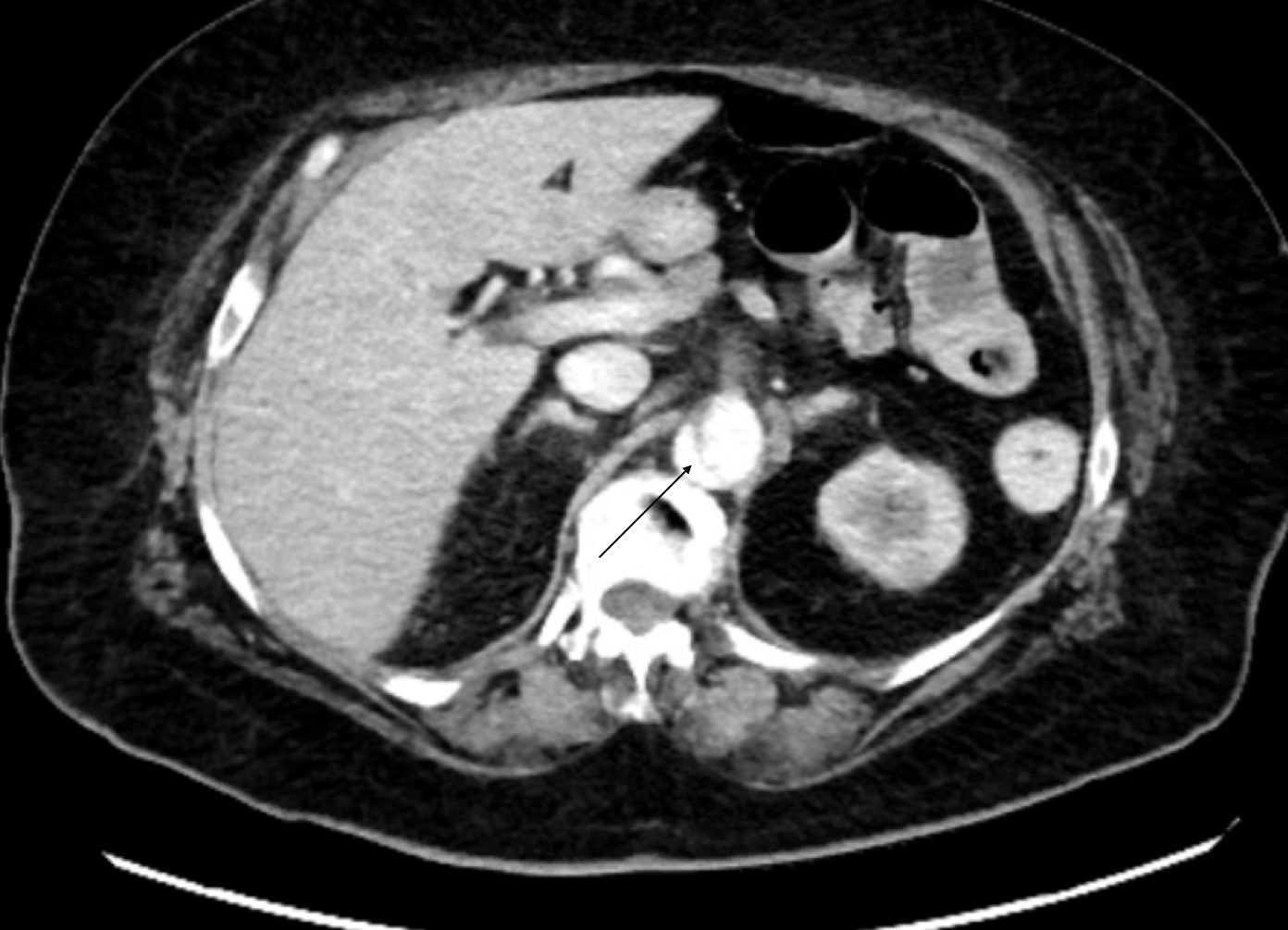
Case Presentation: A 75-year-old female with a past medical history of severe aortic stenosis, paroxysmal atrial fibrillation, hypertension, hyperlipidemia, chronic obstructive pulmonary disease, and morbid obesity presented to the emergency room with dyspnea. She was an active smoker with a 27 pack-year history. Echocardiography revealed a mean transvalvular pressure gradient of 36.1 mmHg and an aortic valve area of 0.89 cm2. She underwent successful transcatheter aortic valve replacement (TAVR) through the left femoral artery with a post-TAVR gradient of 8 mmHg. Severe tortuosity of the descending aorta was noted in the operative report. On post-op day (POD) 0, the patient endorsed nausea and one episode of emesis. Her exam was notable for mild epigastric tenderness and suprapubic fullness. She had a mild leukocytosis to 14,000. On POD 2, she had an additional episode of emesis but no further abdominal pain. Labs were remarkable for WBC 19,000, AST 97, ALT 71 and creatinine of 1.1 from a baseline of 0.5. Infectious work-up, including urinalysis, chest X-ray, right upper quadrant ultrasound, hepatitis panel and HIV serology, was unrevealing. She remained afebrile with blood pressures between 145-190/57-94.On POD 3, she endorsed chills and had a temperature of 99.5 F. Blood cultures were drawn and the patient was started on broad spectrum antibiotics. Her serum lactate was 3.5, WBC increased to 26,000 and her transaminitis worsened with AST 954, and ALT 682.CT scan of the abdomen and pelvis with contrast was performed to rule out cholecystitis and pyelonephritis. It revealed a new aortic dissection extending from the distal thoracic aorta to the level of the bifurcation, involving the celiac trunk and the superior mesenteric artery (SMA).The patient underwent emergent thoracic endovascular aortic repair (TEVAR) with stenting of the SMA. Her hospital course was complicated by cecal ischemia, Desulfovibrio fairfieldensis bacteremia and lower gastrointestinal bleeding requiring multiple transfusions. She was discharged to subacute rehabilitation on hospital day 30 and was well as of her 4-month follow-up visit.
Discussion: TAVR has supplanted open surgical aortic valve replacement as the treatment of choice for severe aortic stenosis, due large part to its lower rate of perioperative complications. Still, as our case demonstrates, TAVR is not without risk. Depending on the institution, post-TAVR patients may be cared for on a hospital medicine service and hospitalists need to be aware of post-procedural complications.This case was an atypical clinical presentation of aortic dissection as a complication of TAVR. Aortic dissection is a life-threatening complication of TAVR with a reported incidence of 0.6% – 1.9%. Valve calcifications and tortuosity of the aorta, as seen in our patient, have been identified as risk factors for vascular complications in transfemoral procedures. Recent studies found the prevalence of severe pain, peripheral pulse deficits, or widened mediastinum on CXR in patients with Type B AD to be 93%, 14%, and 32% respectively. These were notably absent in our patient. Presenting hypertension, as our patient had, was noted to be more prevalent in patients with Type B versus Type A dissection (66% vs 28%) while syncope was less prevalent (18% vs 3%).
Conclusions: As TAVR becomes more common, it is vital for clinicians to maintain a high index of suspicion for aortic dissection and other vascular complications in the post-operative period. Clinical manifestations of aortic dissection may be subtle.