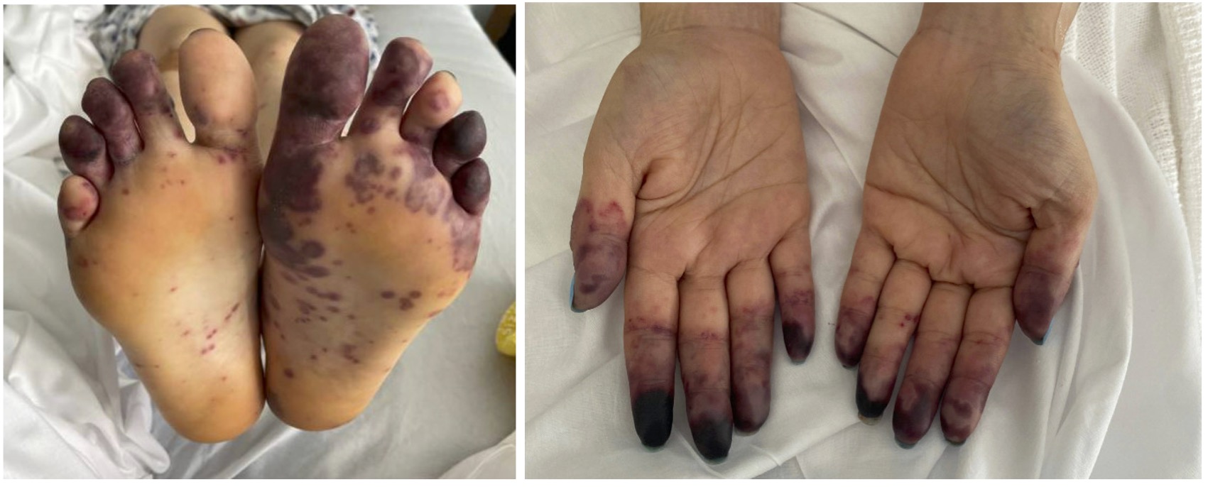
Case Presentation: A 34 year old female presented to the emergency department with several days of rapidly progressive skin and tongue lesions. Over the preceding months, she was evaluated by outpatient otolaryngology for recurrent otalgia and sinusitis, receiving antibiotics and prednisone without improvement. She underwent electromyography with neurology and was diagnosed with peripheral neuropathy of her lower extremities. She also underwent cardiac workup for chest discomfort and was prescribed diltiazem for atrial tachycardia of unclear etiology. Ultimately, she was prompted to present to the hospital due to new skin lesions. Upon arrival, her exam was significant for palpable purpura of her extremities, dusky hand and foot digits (Fig. 1), and a prominent 2 cm tongue ulceration. Her labs were notable for a leukocytosis of 17, platelets of 990, a hemoglobin of 9.4, CRP of 407, ESR of 120, a +PR3 titer >8, a +rheumatoid factor of 43, normal C3/C4, normal ANA, and a subnephrotic range proteinuria. A skin biopsy showed necrotizing small vessel vasculitis with focal granuloma formation. She was diagnosed with granulomatosis with polyangiitis (GPA) with skin, renal, sinus, and nerve involvement. Prompt treatment with pulse dose steroids and rituximab was initiated. Amlodipine, aspirin, sildenafil, nitropaste, pentoxifylline, and plasmapheresis were started for severe digital ischemia. Multiple surgical services assessed the patient’s fingers but were unable to offer intervention. Her course was further complicated by an acute gastrointestinal (GI) bleed requiring transfusion and critical care. Numerous small intestine ulcers were found and attributed to the vasculitis. The bleed stabilized after antiplatelets and anticoagulants were held and with continued rituximab therapy. Her symptoms gradually improved, and she was discharged with close follow-up.
Discussion: GPA is a rare disease, with a frequency of 2-15 per hundred thousand (1). This case highlights the complexities and atypical features of GPA that may impede early diagnosis and lead to fulminant disease. Prior to admission, the patient was seen by specialists as her symptoms evolved. However, her condition progressed rapidly, and she required hospitalization before a unifying diagnosis could be made. Her cardiac findings are nonspecific and not a hallmark of GPA’s illness script; cardiac involvement is estimated to affect less than 44% of GPA patients and most commonly causes pericarditis, but it may also cause arrhythmias due to conduction system involvement (1). In addition, while 30-50% of GPA patients have cutaneous manifestations, typically purpura of the lower extremities, this patient’s digit necrosis was beyond what is typically seen; one retrospective study of 75 GPA cases found only one case of digit necrosis (2). Her fulminant digit ischemia is thus a highlight of her rare presentation. Another unique aspect of her case is the acute GI bleeding. GI tract involvement is thought to occur in the range of 5-11% of GPA cases, with massive bleeds occurring even less often (3).
Conclusions: Granulomatosis with polyangiitis is a rare vasculitis that many providers are aware of; however, early recognition and management of the multifaceted condition remain challenging. Continuing education and maintaining a high index of suspicion, particularly when a patient is having an unusual constellation of clustered symptoms, is imperative for patients with GPA.