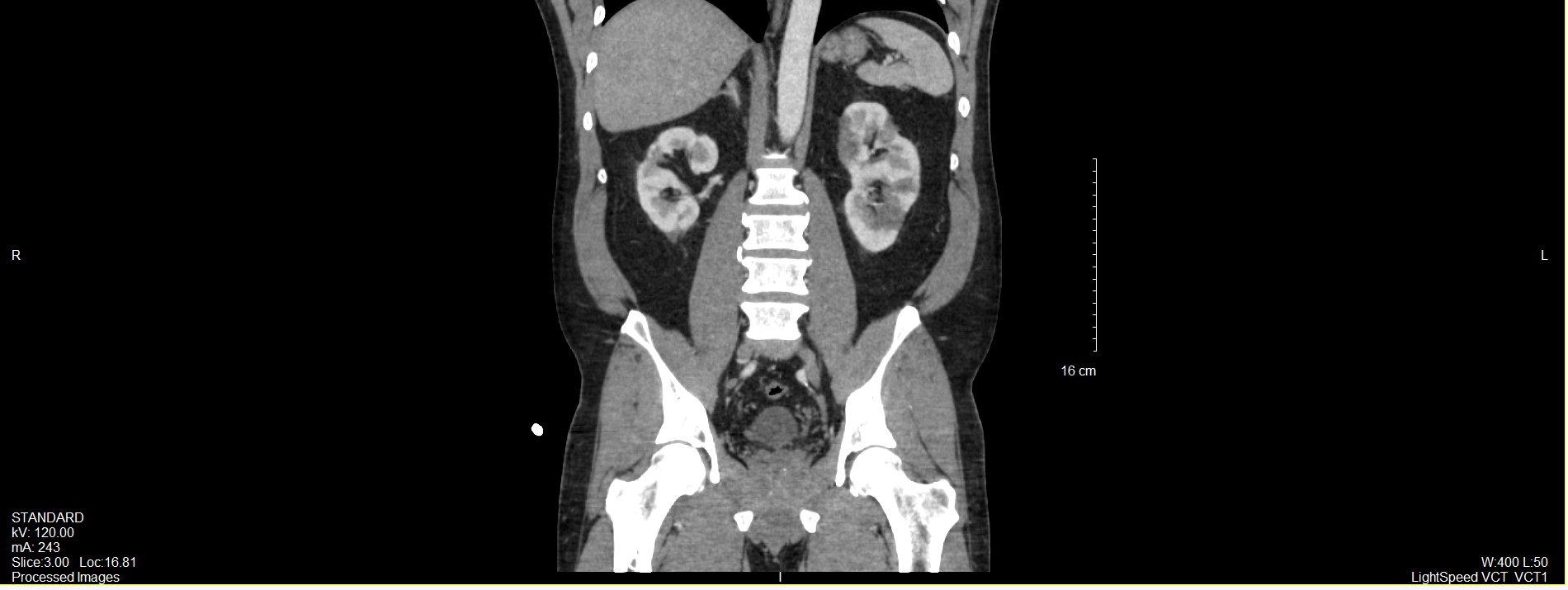
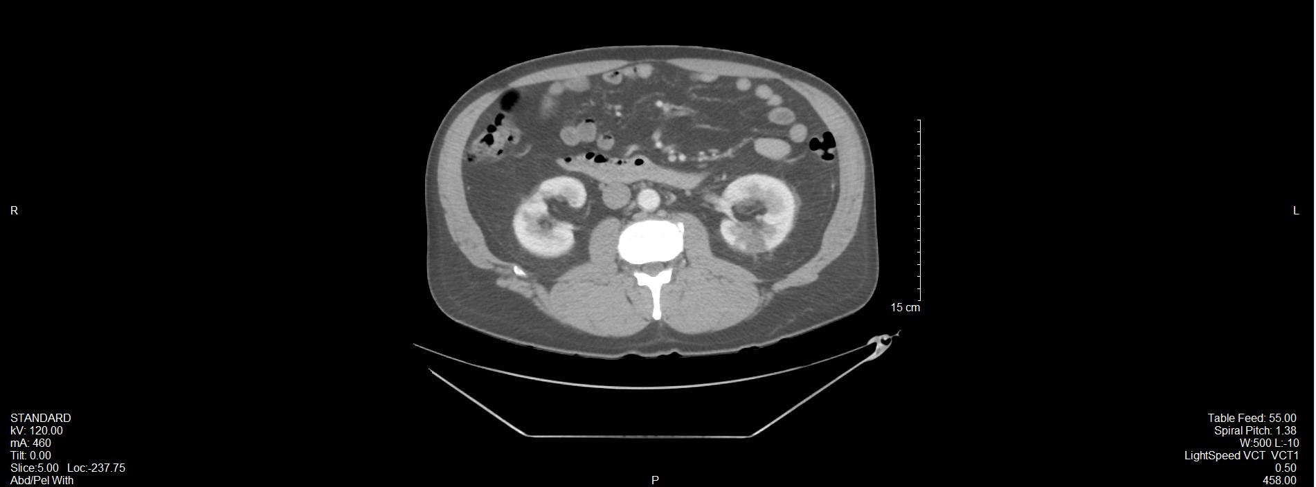
Case Presentation: A 46-year-old male with chronic polycythemia, recurrent DVTs (2015, 2016, 2021), and on long-term testosterone therapy presented with acute chest pain. Troponins were elevated (0.15 → 35.47 → 65.24 ng/mL). EKG showed inferior and lateral STEMI. Labs were notable for hemoglobin 19.5 g/dL, hematocrit 58%. Emergent left heart catheterization demonstrated 100% mid-circumflex occlusion, treated with percutaneous transluminal angioplasty (PTA) and drug-eluting stent (DES) placement. A large left ventricular LV thrombus was noted on echocardiogram. He was started on ticagrelor, therapeutic enoxaparin, and warfarin bridging. Hypercoagulable testing, including JAK2 mutation analysis was negative. Hematology attributed his polycythemia to testosterone therapy and recommended phlebotomy. He was discharged on ticagrelor, therapeutic enoxaparin injections, warfarin for bridging, beta-blockers, and statins.Shortly after discharge, he returned with acute abdominal pain. CT angiography revealed multiple left renal cortical hypodensities consistent with renal infarcts. He was started on heparin, later transitioned back to therapeutic enoxaparin with warfarin bridging, and discharged with close cardiology and hematology follow-up.
Discussion: Testosterone therapy remains controversial regarding cardiovascular safety, particularly when serum levels exceed physiologic ranges. Evidence shows that hematocrit ≥52% significantly increases the risk of major adverse cardiovascular events and venous thromboembolism. Case reports and observational studies have linked anabolic steroids and high-dose testosterone with venous thromboembolism and myocardial infarction, while renal infarction is rarely described. Our case of acute STEMI, left ventricular thrombus, and renal infarction in a patient using high-dose testosterone complicated by chronic polycythemia highlights the severe thrombotic potential of supraphysiologic androgen exposure. Erythrocytosis is the most common adverse effect of testosterone therapy, that increases blood viscosity and contributes to hypercoagulability. In this patient, long-term high-dose testosterone created a persistent prothrombotic state that likely precipitated coronary thrombosis, LV thrombus formation, and subsequent renal infarction despite appropriate antiplatelet and anticoagulant therapy. This case underscores essential principles: clinicians must screen for non-prescribed anabolic steroid use, and short-acting injectable formulations should be recognized as carrying the highest risk of erythrocytosis.
Conclusions: This case demonstrates the increased risk of both venous and arterial thrombosis associated with testosterone-induced polycythemia. While physiologic testosterone replacement may be safe in appropriately selected patients, supraphysiologic dosing markedly elevates the risk of erythrocytosis, hyperviscosity, and end-organ ischemia. Prospective studies correlating hematocrit changes with infarction risk are lacking, and renal infarction remains rarely reported in this context. Clinicians should closely monitor hematologic parameters, assess cardiovascular risk, and counsel patients on the dangers of excessive testosterone exposure. Testosterone therapy should be reserved for those with clear clinical and biochemical hypogonadism, with routine follow-up to prevent avoidable complications.