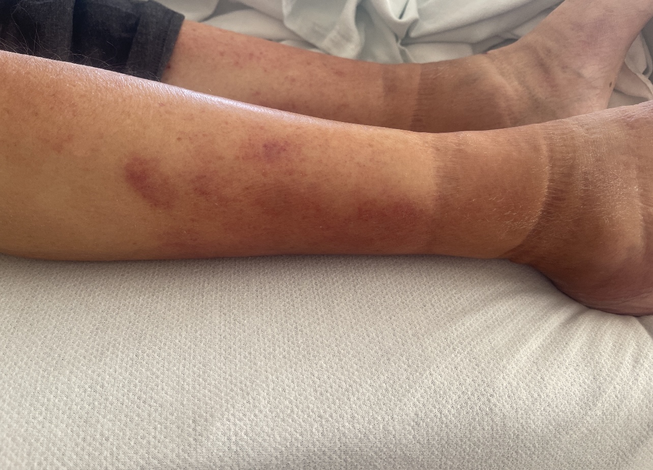
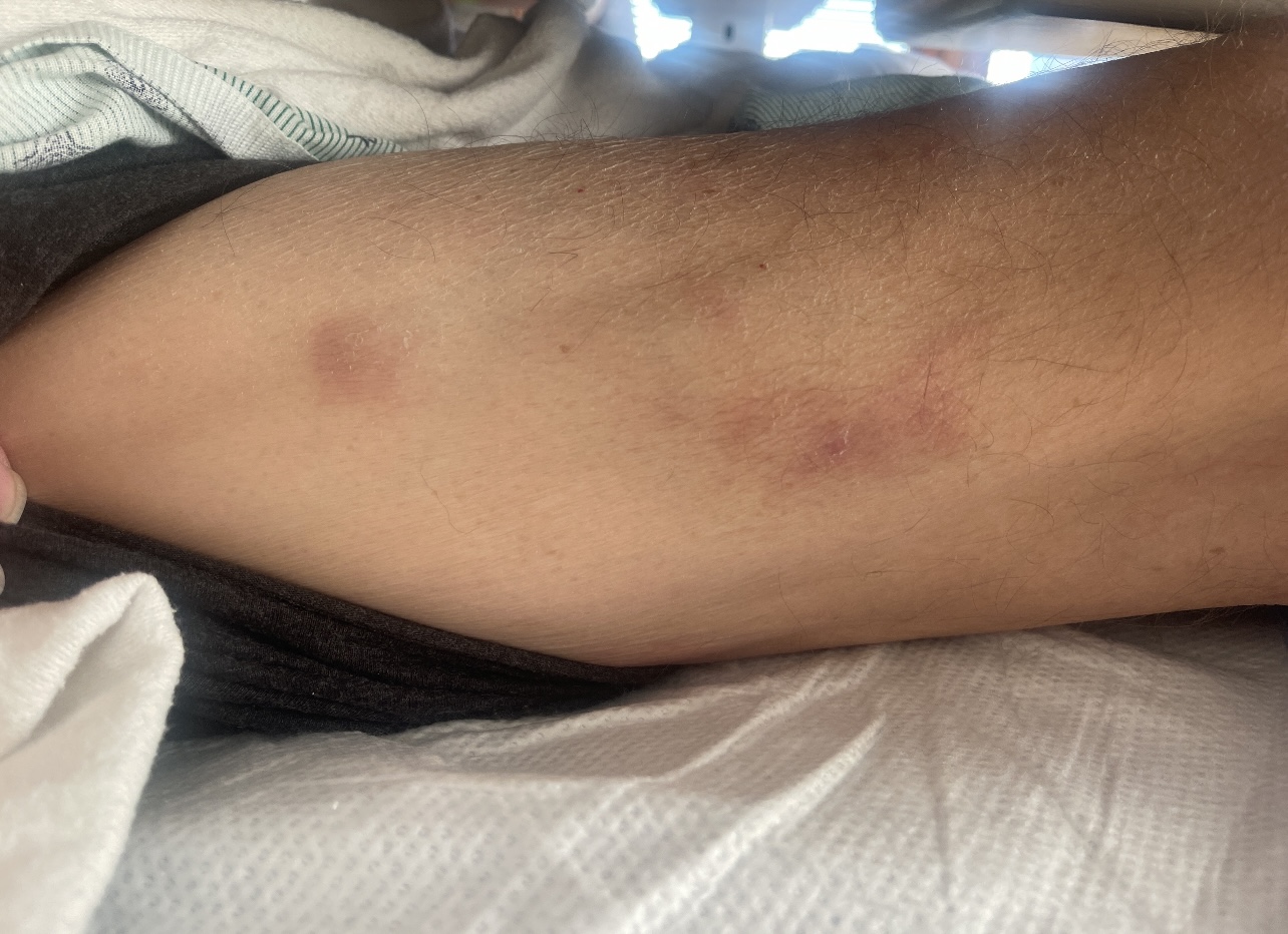
Case Presentation: A 63-year-old male with a past medical history of end-stage renal disease status post living-related kidney transplant three years ago presented with dyspnea and joint pain. His immunosuppressive regimen included mycophenolate mofetil, prednisone, and belatacept, which was switched to everolimus due to concern for synovitis at outpatient rheumatology one month prior when he was noted with erythematous nodules on lower extremities (BLE).On exam, patient appeared ill with dry mucous membranes, but was alert, oriented, and afebrile. On dermatological evaluation, hands were swollen and BLE had pitting edema, along with pruritic, erythematous, firm, and indurated macules and subcutaneous nodules. TB and viral panels were negative, except for EBV titer, elevated at < 500. Fungal culture was negative, except for elevated histoplasma urine antigen of 0.44 ng/mL. Patient tested positive for RSV bronchiolitis, likely the cause of his dyspnea.Skin biopsies and tissue cultures of the nodules were positive for 4+ acid fast bacilli (AFB). Steroids were tapered to 5mg. No antibiotics were initiated, awaiting biopsy culture results of mycobacterium. Patient was discharged on ribavirin for RSV. Ribavirin was discontinued days later due to elevated liver enzymes, lactic acidosis, and respiratory alkalosis. Patient was re-admitted three weeks post admission with weakness, melena, and decreased appetite. On exam, subcutaneous nodules had extended to his back, chest, and face. In addition to electrolyte and hematologic abnormalities, EBV titers had increased to 14,096. Previous skin biopsies with PCR identified Mycobacterium haemophilum. Patient was started on azithromycin 500mg, rifabutin 300mg daily, and ciprofloxacin 500mg BID.
Discussion: Subcutaneous nodular findings are associated with numerous causes, including erythema nodosum, other panniculitis, medium vessel vasculitis, mycobacteria and fungal infections, thereby delaying diagnosis. In immunosuppressed patients, nontuberculous mycobacteria often manifests as pulmonary disease, followed by skin and soft tissue infection [1]. A thorough workup is essential to rule out underlying pathologies, though this may delay treatment. Treatment depends on the species and is complex, requiring considerations for drug toxicities, long-term multidrug regimens, and drug-drug interactions [2]. For fastidious, difficult-to-isolate pathogens like Mycobacterium haemophilum, NAAT may be vital in instances where tissue culture resolution can take weeks. In a similar case involving a transplant recipient, NAAT revealed Mycobacterium haemophilum though tissue cultures were negative, allowing prompt diagnosis and treatment [3].
Conclusions: Mycobacterium haemophilum is rare, but can have cutaneous manifestations in patients with high dose immunosuppressive requirements. Our patient’s antibiotic coverage was delayed as the culture takes 4 to 6 weeks. The aim of this case report is two-tiered: 1) to raise awareness amongst clinicians about dermatological findings in the transplant population, and 2) to encourage clinicians to reference literature for workup for underlying causes to have better treatment outcomes and prevent progression of conditions.