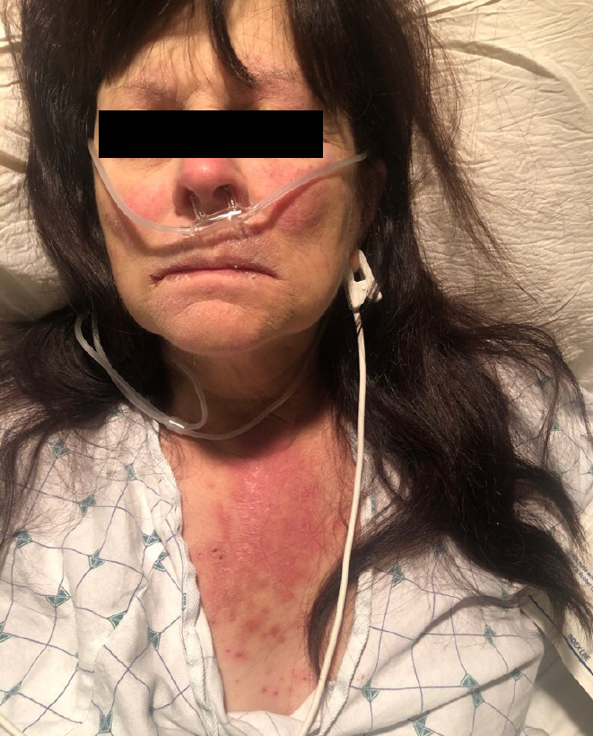
Case Presentation: A 63-year-old woman with multiple sclerosis, hypertension, and venous insufficiency was admitted with shock and diffuse rash. Two months earlier, she had a confluent, erythematous, pruritic, painful, bilateral foot rash that spread from the dorsi to the ankles with associated swelling. Clotrimazole resulted in mild improvement. Two weeks prior to admission, she developed V-shaped chest erythema and plaques, pustules with overlying scale on her arms, thighs, abdomen, and back, and a malar rash. She noted fatigue, anorexia, and decreased urine output. On admission, she quickly decompensated with hypotension refractory to fluids requiring vasopressor support and received broad-spectrum antibiotics.Laboratories showed a normal WBC count, creatinine of 11 mg/dL (baseline 0.8), urinalysis revealing ATN, ANA titer 1:80, negative anti-dsDNA, anti-SSA/SSB, anti-RNP, and anti-smooth muscle antibodies, negative ANCAs, and normal C3, C4, and creatine kinase. Lower extremity skin swabs were negative for HSV, fungi, and AFB, and grew MSSA, which was treated with mupirocin. Biopsy showed subacute spongiotic dermatitis.Drug reactions were considered, particularly to hydrochlorothiazide and furosemide, but she had taken those medications for months prior to symptom onset, lowering suspicion for these agents as the etiology.On further questioning, the patient’s partner noted exposures to doxycycline prescribed for cellulitis. After the first exposure one year prior, she developed a diffuse rash that was ascribed to a concurrent antibiotic. The second exposure, which was not documented in the EMR, directly preceded the onset of the current diffuse rash. She was still taking doxycycline just prior to admission.Based on the history, exam, and biopsy, a diagnosis of acute photodistributed exanthematous pustulosis (APEP), a rare form of acute generalized exanthematous pustulosis (AGEP), was made. Doxycycline, the likely culprit, was immediately added to the allergy list.Blood cultures remained sterile, and antibiotics were stopped within four days. Her condition improved with supportive care, and the rash cleared dramatically with high-potency topical steroids.
Discussion: While the initial foot rash was likely tinea pedis, venous insufficiency, and possible superimposed bacterial cellulitis, the diffuse rash and shock suggested a separate process. The differential diagnosis of shock with diffuse rash includes autoimmune, infectious, toxin-mediated, and drug-induced etiologies. Antibiotics are reasonable in unstable patients until infection is ruled out. Rash morphology and distribution, laboratories, biopsy, and importantly, a detailed history, help narrow the differential. Autoimmune considerations include dermatomyositis, subacute cutaneous lupus erythematosus (with or without systemic lupus), and drug-induced lupus. Infectious considerations include toxic shock and scalded skin syndrome. Drug reactions such as SJS/TEN, DRESS/DIHS, or AGEP should be suspected with medication exposures, particularly sulfonamide diuretics or antibiotics. AGEP occurs hours to days after exposure and quickly resolves after drug discontinuation. Severe cases may include SIRS or shock. Steroids are recommended for severe cases and are typically given topically.
Conclusions: Hospitalists should be aware of the broad differential for shock with diffuse rash. A thorough medication history may identify key exposures not documented in the EMR.