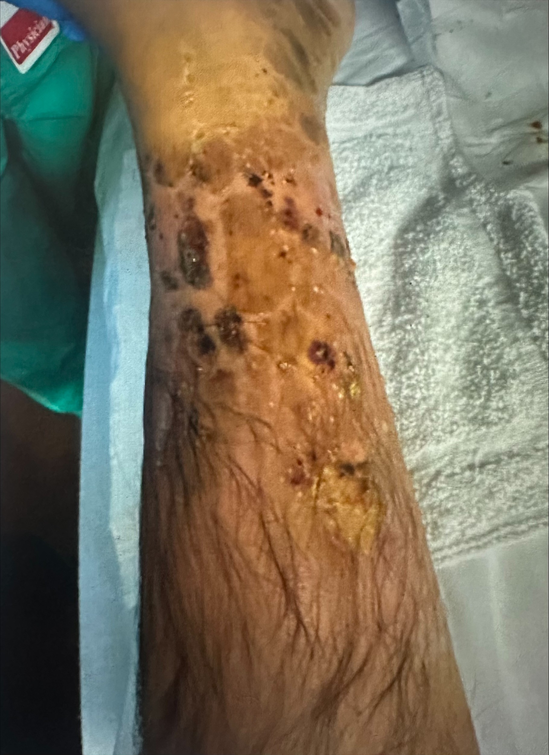
Case Presentation: A 38-year-old man with a 17-year history of well-controlled ulcerative colitis (UC) and rheumatoid arthritis presented with more than ten episodes of hematochezia, severe abdominal cramping, and several weeks of progressive left lower extremity swelling, erythema, and purulent drainage. On admission, he was febrile and tachycardic. Examination showed left lower extremity edema with scattered purulent ulcerations and marked tenderness out of proportion to exam.Labs showed WBC 14.0 K/µL, hemoglobin 8.1 g/dL, CRP 25.1 mg/dL, and a markedly elevated fecal calprotectin of 3,440 µg/g. A gastrointestinal pathogen panel was negative. Broad-spectrum antibiotics were started for presumed severe skin and soft tissue infection, but he showed minimal improvement. Given the lack of response, MRI of the leg was obtained and demonstrated skin thickening, subcutaneous edema, and myositis without abscess, gas, or osteomyelitis. A punch biopsy of an ulcer revealed a neutrophilic infiltrate with suppuration and no microorganisms. Flexible sigmoidoscopy confirmed a severe UC flare with circumferential colitis, friability, and deep ulcerations. High-dose intravenous corticosteroids were initiated, resulting in rapid improvement in hematochezia, abdominal pain, and—critically—marked improvement in left leg pain, edema, and wound appearance. Antibiotics were discontinued once infection had been excluded and biopsy supported a neutrophilic dermatosis, confirming pyoderma gangrenosum (PG) in the setting of an active UC flare.
Discussion: PG is a rare neutrophilic dermatosis occurring in approximately 0.75–1% of patients with ulcerative colitis. Although often appearing near the time of UC diagnosis, PG may occur decades later. Early PG is difficult to recognize because it lacks the classic undermined violaceous borders and necrotic base, instead presenting as tender pustules or nodules that closely mimic cellulitis. Pain out of proportion to exam is a useful early clue. PG may extend to fascia, muscle, or joints, mimicking necrotizing fasciitis, myositis, or osteomyelitis. Imaging helps distinguish PG from infection, while biopsy typically shows a sterile neutrophilic infiltrate. As a diagnosis of exclusion, PG should only be confirmed once infection and other mimics have been ruled out.Management focuses on treating the underlying UC flare and initiating immunosuppression. Systemic corticosteroids or cyclosporine are first-line therapies, though anti-TNF agents have shown high rates of healing in PG associated with inflammatory bowel disease.
Conclusions: This case highlights the importance of maintaining a high index of suspicion for early-stage pyoderma gangrenosum in patients with UC who develop painful skin lesions lacking classic necrotic or violaceous features—even many years after initial diagnosis. PG can closely mimic severe skin and soft tissue infection and may prompt broad-spectrum antibiotics or invasive evaluations when not considered early. For hospitalists, timely recognition of PG, particularly when associated with an active UC flare, is essential to avoid delays in immunosuppressive therapy and prevent superimposed infection, scarring, or limb-threatening complications. Prompt initiation of systemic corticosteroids targeting the underlying flare leads to rapid clinical improvement and is critical for optimal outcomes.