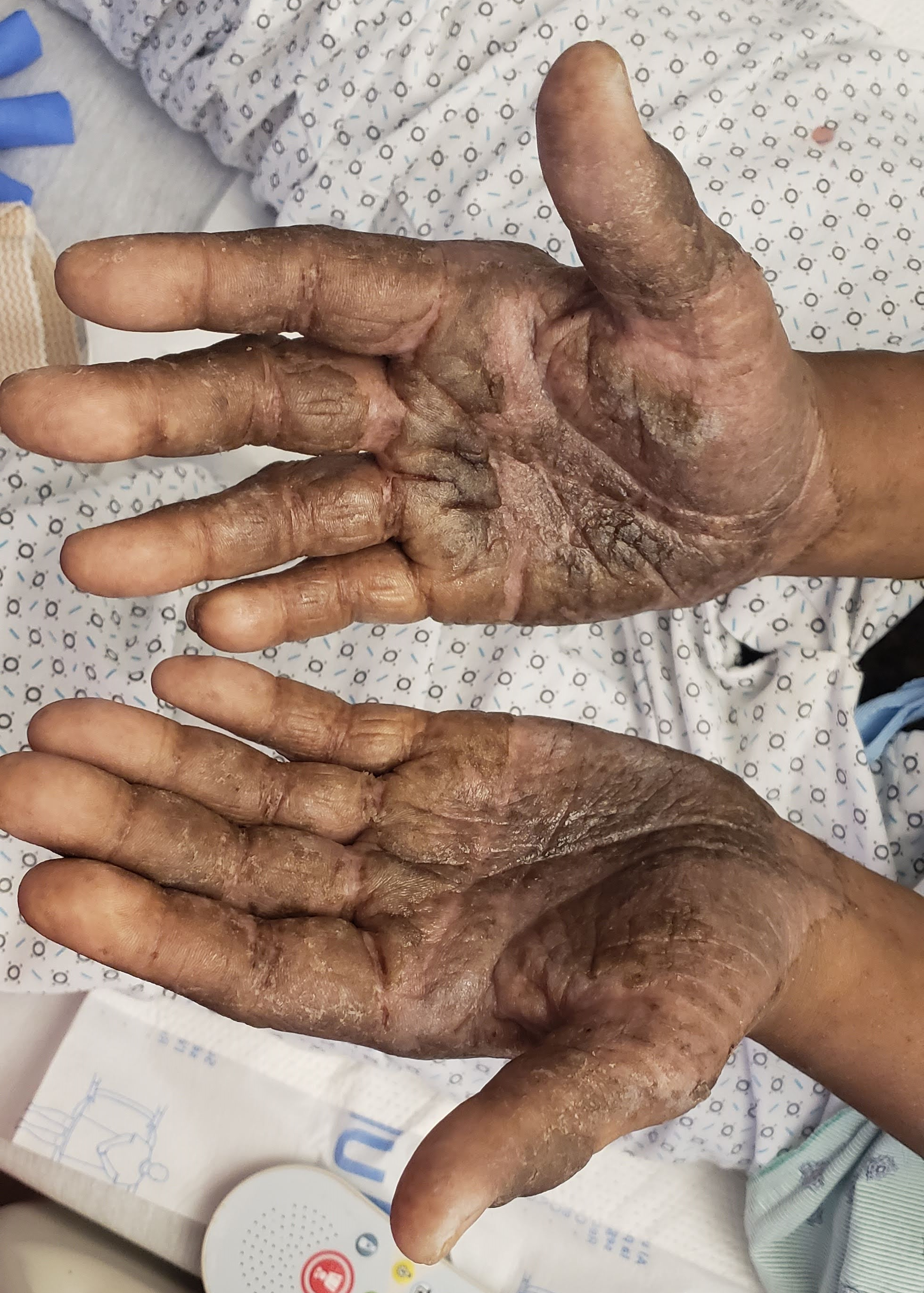
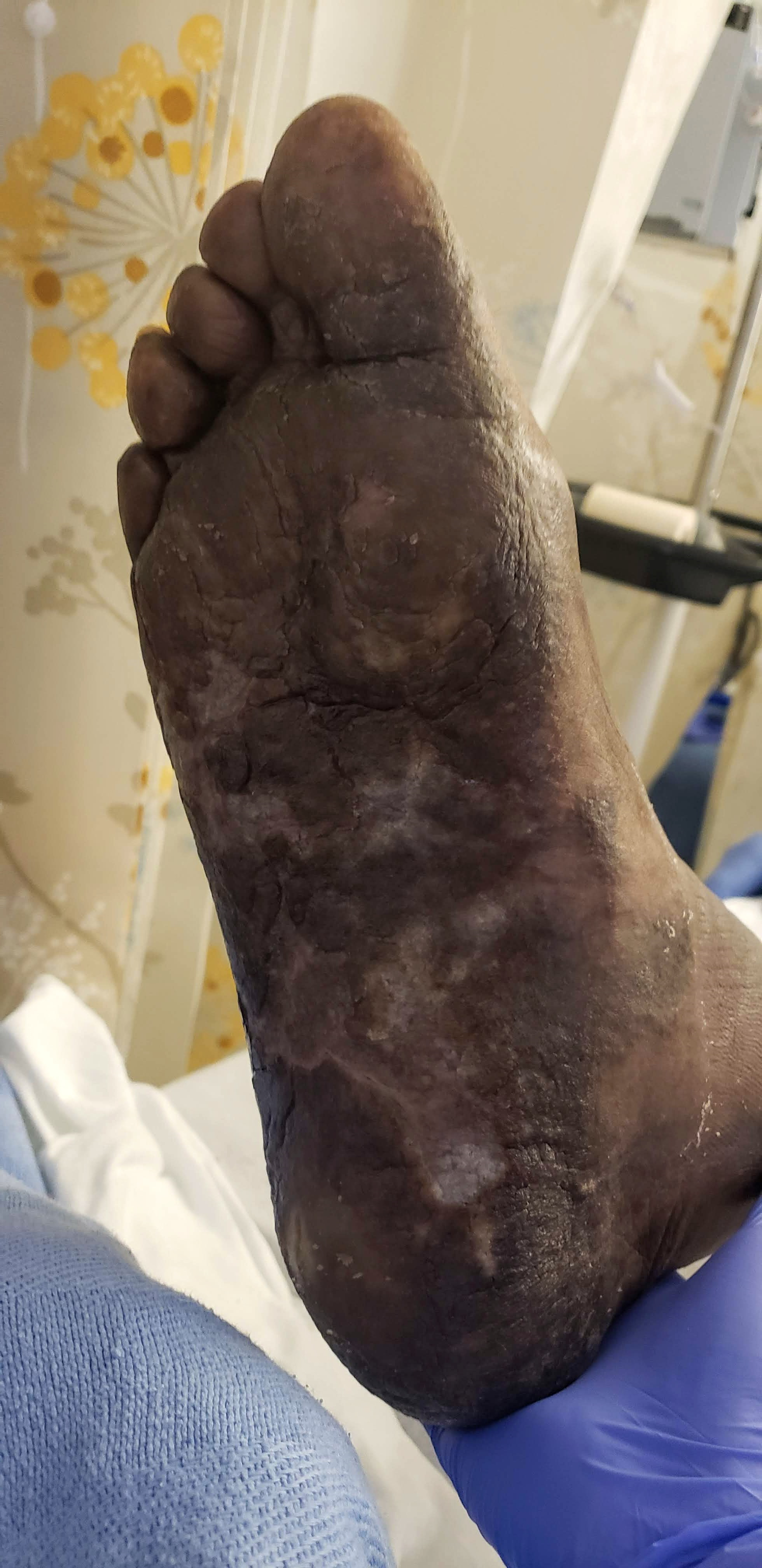
Case Presentation: A 74 year old man with a history of hypertension and chronic hepatitis C infection presented to the hospital with a worsening pruritic rash on his palms and soles. The rash developed acutely one year prior and biopsy was suggestive of eczematous dermatitis. Symptoms persisted in spite of multiple treatments including topical and systemic corticosteroids, phototherapy, and tralokinumab, a human IgG4 monoclonal antibody. He denied a childhood history of atopy or allergies and had no prior history of atopic dermatitis. In the preceding two weeks, he noted nightly subjective fevers and an unintentional weight loss of ten pounds. On admission to the hospital, his temperature was 39C, blood pressure was 120/72, and pulse was 113. He had a hyperpigmented, hyperkeratotic rash with fissuring involving the palms and soles and an overlying cellulitis of the left lower extremity. His white blood cell count was 34.3 K/MM3 with 7.1% peripheral blasts, hemoglobin was 7.9 g/dL, and platelets were 78 K/MM3. A bone marrow biopsy was consistent with acute myeloid leukemia. He was started on azacitidine and venetoclax. One month later, his rash had nearly completely resolved.
Discussion: Adult onset atopic dermatitis is less commonly seen than persistence or recurrence of atopic dermatitis from childhood. In patients with atypical features or refractory disease, it may be a manifestation of another underlying condition. Some neoplastic diseases have been known to trigger cutaneous manifestations and an underlying malignancy may have been the driver for this patient’s cutaneous changes. On review, the patient had a new pancytopenia when he initially presented for his rash with a white blood cell count of 3.4 K/MM3, hemoglobin of 7.8 g/dL, and platelets of 118 K/MM3. Five months later, he was admitted for an abscess. At that time, white blood cell count was 1.5 K/MM3, absolute neutrophil count was 0.4 K/MM3, hemoglobin was 7.8 g/dL, and platelets were 97 K/MM3. His pancytopenia was attributed to untreated hepatitis C infection and active infection so further workup was deferred to the outpatient setting. Although rare, he may have had an evolving bone marrow process that precipitated his initial rash. After treatment for acute myeloid leukemia was started, his rash rapidly improved with near complete resolution in a month, further supporting a connection between his treatment resistant eczematous dermatitis and acute myeloid leukemia.
Conclusions: Severe treatment resistance should prompt re-evaluation, particularly in the context of other abnormalities. Similar to looking for secondary causes of treatment resistant hypertension, physicians should also consider potential underlying causes when evaluating treatment resistant skin conditions. Many patients present to the hospital for pieces of their care and emphasizing key follow-up features to ensure smooth transitions of care between the inpatient and outpatient setting can improve patient outcomes. For this patient, this may have prompted an earlier work up for the pancytopenia and profound neutropenia. A patient’s presentation to the hospital can also be a good opportunity to consider the clinical picture as a whole. In this case, the rapid onset of severe, treatment-resistant symptoms and concomitant new pancytopenia may have been clues that an underlying process was driving his skin changes.