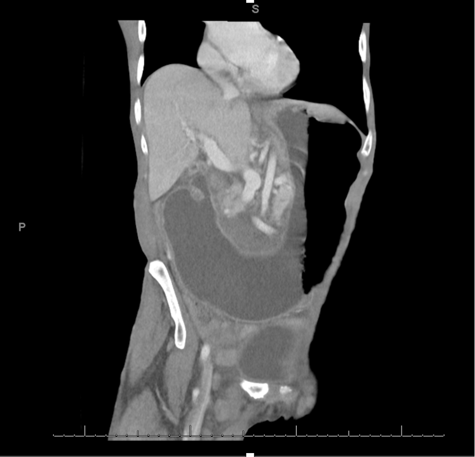
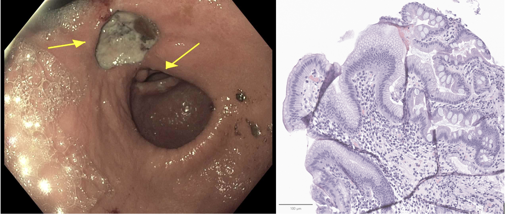
Case Presentation: A 65-year-old woman with a history of opioid use disorder presented to the emergency department with abdominal pain and vomiting. Without her methadone, she was in withdrawal and vitals were notable for tachycardia (101 bpm) and hypertension (184/105 mmHg). History was limited by anxiety and discomfort. Physical examination revealed cachexia with a Body Mass Index of 12.73 kg/m2, edentulism, and left upper quadrant abdominal tenderness. Labs were notable for an anion gap of 20, and beta-hydroxybutyrate of 1.29 mmol/L. Computed Tomography of the abdomen showed massive gastric distension concerning for gastric outlet obstruction (Figure 1) and subsequent nasogastric tube placement had coffee-ground output. After initial stabilization, she underwent esophagogastroduodenoscopy (EGD) which revealed a 2.5 cm cratered ulcer compressing the pylorus and obstructing the gastric outlet (Figure 2). Biopsy revealed H. pylori gastritis with focal intestinal metaplasia (Figure 2).Her quadruple therapy course was complicated by nausea, abdominal pain, and diarrhea. Clarithromycin-based treatment was avoided due to QT prolongation and need for methadone.She was started on tube feeds for her malnutrition via NG tube, but her uninsured status precluded discharge with tube feeds. She remained admitted until she could meet her calorie needs by mouth and was discharged with one week remaining of quadruple therapy. She was readmitted with GOO less than two months later and repeat EGD redemonstrated the ulcer. Surgery was consulted for consideration of partial gastrectomy, but she was discharged home for nutritional rehabilitation before operative management. She was found dead at home less than a month after discharge.
Discussion: Peptic ulcer disease (PUD) affects approximately 3.4 million (1%) in the US population and is typically caused by Helicobacter pylori infection (42%) or aspirin and NSAID use (36%). Twenty to twenty-five percent of patients with PUD experience complications, most commonly bleeding (73%) or perforation (9%), however, in 2% to 3% of cases, may cause gastric outlet obstruction (GOO).This patient with severe protein calorie malnutrition from gastric outlet obstruction exemplifies a rare complication of H. pylori associated peptic ulcer disease. The pathogenesis of obstruction involves edema and spasm combined with chronic fibrosis. Treatment of H. pylori infection reduces inflammation, relieving obstruction. A small study showed resolution of symptoms and endoscopic evidence of ulcer resolution in 17/20 patients within 2 months of treatment, and most other studies show resolution after medical therapy. Recurrence of obstruction (as seen in this case) is rare.In refractory cases, endoscopic balloon dilation or gastrectomy may be considered. There is little data available regarding the frequency of procedural management, but 22% to 67% of patients may require serial dilation and 8% to 51% may require operative management.
Conclusions: H. pylori associated gastric outlet obstruction is rare, and multiple factors contributed to the complexity of this case. QTc prolongation with methadone use limited use of ondansetron or a clarithromycin-based antimicrobial regimen. Excess side effects from her regimen may have decreased medication adherence after discharge. Additionally, the patient’s uninsured status may have contributed to a late presentation, prolonged inpatient stay, readmission, and death.