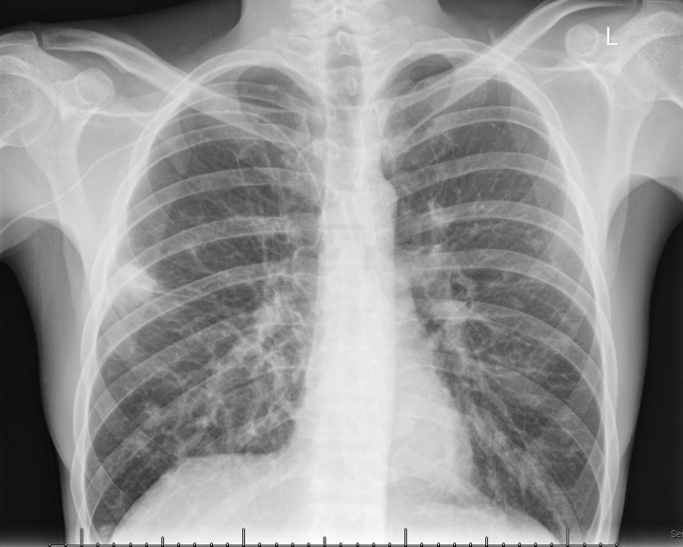
Case Presentation: This case underscores the diagnostic challenge of pulmonary Kaposi’s Sarcoma (KS) in HIV/AIDS patients, emphasizing oral mucosal lesions as significant markers, especially alongside biopsy-proven results when bronchoscopy findings are negative.A 36-year-old HIV/AIDS male ([CD4 3 cells/mm³, viral load 1.4 million copies/mL]), with a history of cryptococcal meningitis, presented with a 3-week history of fever, pleuritic chest pain, dyspnea, and scant hemoptysis. Socioeconomic barriers led to discontinuation of antiretroviral therapy (ART) and prophylactic medications. One month earlier, he was hospitalized for pneumonia at an external facility, receiving an unknown antibiotic regimen.On presentation, he appeared cachectic, in severe respiratory distress, with diminished breath sounds and scattered rhonchi. Violaceous plaques were noted on the hard palate and extremities, with an 8-month progression. Laboratory investigations revealed hemoglobin 8.7 g/dL, MCV 67 fL, and elevated LDH (298 unit/L) and CRP (4.2 mg/dL). CT chest showed bilateral multifocal opacities with flame-shaped morphology, surrounding ground-glass opacities, and interlobular septal thickening.The patient required high-flow nasal cannula for acute hypoxemic respiratory failure and was treated empirically with Vancomycin, Cefepime, and Flagyl for presumed pneumonia but showed limited improvement after 7 days. Bronchoscopy excluded endobronchial lesions, and BAL failed to identify microorganisms, excluding infectious etiologies. Positive EBV IgG and elevated HHV-8 DNA PCR (9,100 copies/mL), along with immunolabeling of HHV-8 in skin lesion biopsies and BAL samples, confirmed KS. Negative results for tuberculosis and cryptococcosis helped avoid immune reconstitution inflammatory syndrome (IRIS).The patient was reinitiated on ART (Biktarvy), PJP prophylaxis with Bactrim, and fluconazole for cryptococcal meningitis maintenance. He responded well to doxorubicin chemotherapy, with resolution of pulmonary opacities and skin lesions after three of six cycles, allowing oxygen therapy de-escalation. Long-term management and surveillance by Special Immunology and Oncology teams are ongoing.
Discussion: Kaposi’s Sarcoma, a vascular neoplasm linked to HHV-8, poses diagnostic challenges in HIV/AIDS patients, particularly pulmonary KS, which often accompanies mucocutaneous disease (80-90% of cases). In this instance, violaceous mucocutaneous lesions, in conjunction with radiographic patterns, prompted consideration of pulmonary KS. The flame sign pattern, explained by inflammation, vascular proliferation, and vasodilation, noted with surrounding ground-glass opacities, are characteristic of pulmonary KS. While bronchoscopy often reveals pathognomonic mucosal lesions, their absence does not exclude the diagnosis. Confirmation was achieved through positive HHV-8 serology and BAL sampling, corroborated by skin biopsy results. Deferred gastrointestinal evaluation limited our ability to exclude potential involvement, despite suggestive findings on fecal occult blood and iron deficiency anemia. ART remains the cornerstone of treatment, and pegylated liposomal doxorubicin is a preferred first-line option for visceral involvement, demonstrating efficacy in our patient.
Conclusions: Mucocutaneous assessment and radiographic findings are critical for identifying pulmonary KS in HIV/AIDS patients, particularly those with poorly controlled disease.