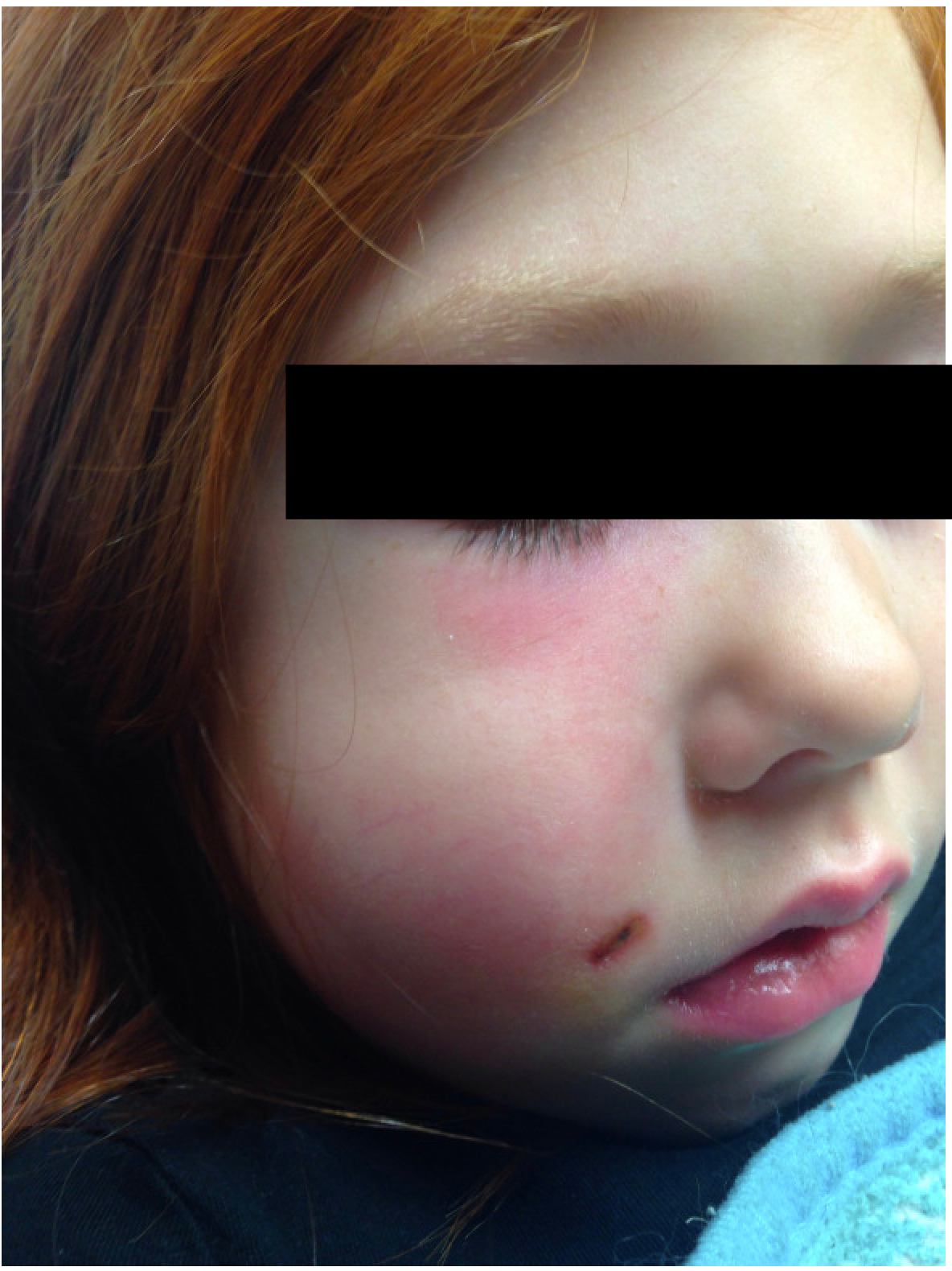
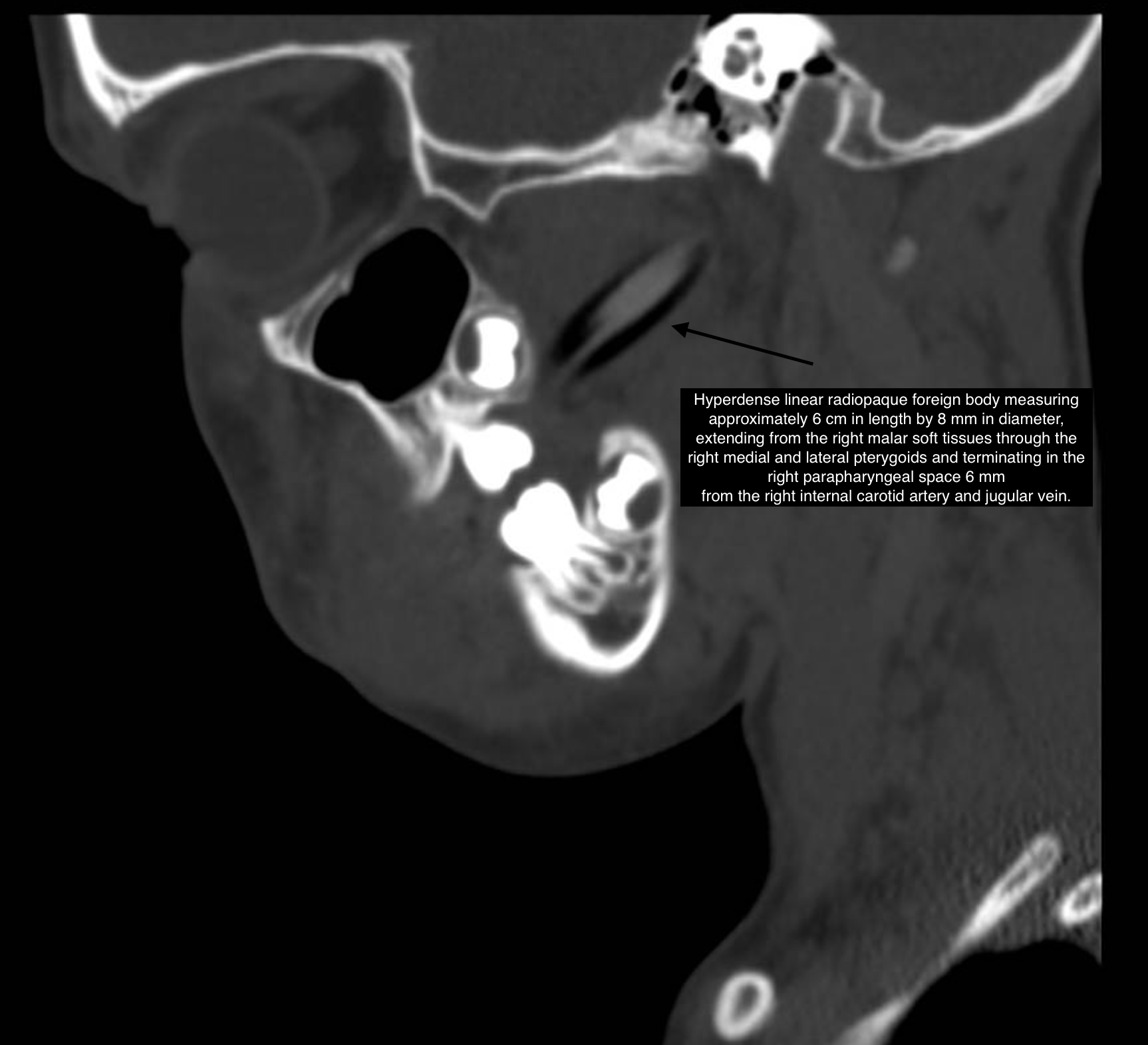
Case Presentation: A 5-year-old healthy female presented to the Emergency Department (ED) three times within 48 hours for a right facial wound with worsening swelling and redness. Prior to presentation, the patient was running down the hall with a colored pencil when she collided with her father. Half of the broken pencil was located but the other half was unable to be found in the house. The parents suspected that it was lodged inside her cheek. The patient was taken to a level I trauma center ED, where a facial bone xray and ultrasound (US) were obtained. Xray noted a linear opacity projecting over the right ramus extending posteriorly through the oropharynx. US revealed a 0.4 x 0.4 x 0.1 cm foreign body (FB) near the facial wound. Pediatric Otolaryngology was consulted. They did not feel the imaging or physical exam were consistent with a retained FB and recommended discharge with follow-up. The patient was brought to a second ED 10 hours after discharge for further evaluation. Images from the first ED were reviewed and the facial trauma service was consulted. They recommended topical polysporin ointment to the facial wound, discharge home and outpatient follow-up. The patient returned to the ED the following day due to worsening redness, cheek swelling, and inability to open her mouth. Facial trauma service was reconsulted and recommended repeat ultrasound, which was negative for FB. They noted a firm area in her right upper buccal sulcus with a differential of edema versus foreign body. The patient was admitted for IV hydration, ampicillin-sulbactam IV, and monitoring for presumed cellulitis. Her facial swelling worsened overnight and parents echoed their concern for a retained pencil fragment. A maxillofacial CT was obtained, revealing a 6 cm x 8 mm FB which terminated in the right parapharyngeal space 6 mm from the right internal carotid artery and jugular vein. The patient was taken to the operating room for surgical resection by plastic surgery and the FB was identified as the missing colored pencil.
Discussion: We present a case of a large, retained FB, located millimeters from the carotid artery and jugular vein, whose identification and removal was delayed despite appropriate initial diagnostic modalities and subspecialty consultations. While US is a highly trusted modality for quick, cost-effective and radiation-sparing imaging, especially in children, it is important to remember that it has a wide range of sensitivity and specificity. For FB detection, sensitivity may be as low as 50% or as high as 90%, in large part due to operator skill. If FB is identified, US has a specificity for materials including wood that may be as high as 70-97%. The patient’s US was performed in a level 1 trauma center by a presumably highly skilled US operator and failed to diagnose a large FB. Thus, it is likely there are other factors besides operator error contributing to poor sensitivity. This case highlights the pitfalls of being overly confident in US to rule out foreign bodies in patients with a compelling history and concerning physical exam. This case also demonstrates the ongoing struggle that ensues as we weigh the benefits of advanced imaging like CT with the risks of radiation exposure in young children.
Conclusions: This is a unique case of a retained FB that eluded multiple evaluations. This case demonstrates the limitations of US in ruling out a foreign body and highlights the importance of further investigation if the clinical picture does not correlate with the working diagnosis.