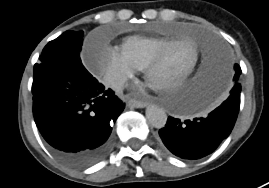
Case Presentation: A 69-year-old woman with stage IV breast cancer presented with new onset abdominal pain, dyspnea on exertion, and fatigue causing her to present to our hospital. She denies chest pain currently but admits some in past 2 weeks. Denies palpitations, orthopnea, fevers, chills, recent illness, or exposures. CT abdomen showed an incompletely visualized pericardial effusion plus ascites. Echocardiogram further defined the large circumferential pericardial effusion with evidence of early cardiac tamponade including right atrial systolic collapse, focal strands, dilated IVC and hepatic veins. Physical exam was positive for Becks triad (decreased blood pressure, increased jugular venous pressure, and muffled heart sounds). Vital signs revealed heart rate of 110s, tachypneic to the 30s, afebrile, hypotensive with systolic blood pressure in 90s and saturating 96% on room air. EKG showed sinus tachycardia. Pertinent labs included hyponatremia with elevated lactic acid to 3.27, leukocytosis with a WBC of 16.5, troponins were within normal limits. Pericardiocentesis revealed a large hemorrhagic pericardial effusion draining over 900 mL. Pericardial fluid analysis of the exudative fluid consisted predominately of red blood cells. The sample did not show evidence of malignant cells. The drain output tapered off over several days with resolution of her pericardial effusion. Her pericardial drain was removed, and she was discharged on NSAIDs and colchicine with cardiology follow up. The final diagnosis being idiopathic pericarditis leading to a large hemorrhagic pericardial effusion.
Discussion: Pericardial effusion is a relatively common syndrome and has a diverse set of etiologies including infections, malignancy, radiation/iatrogenic, pericardial injury, metabolic disturbances, endocrine disease, connective tissue disease, autoimmune disorders, trauma, or idiopathic. Pericardial effusion presentation is a large spectrum that can vary from asymptomatic to cardiac tamponade. Pericardial drainage is recommended in most large pericardial effusions or those causing cardiac tamponade. Pericardial fluid can be either purulent, serous, serosanguinous, or hemorrhagic. In the case of hemorrhagic effusion, an etiology of malignancy, iatrogenic, and tuberculosis (in endemic regions) become more likely, however the possibilities remain broad. Large hemorrhagic pericardial effusions resulting from idiopathic (presumed viral) pericarditis are rare but possible.
Conclusions: Hemorrhagic pericardial effusions are often the result of malignancy, tuberculosis, or iatrogenic; but viral etiologies should also be considered, as was the case with our patient. Hemorrhagic effusions need aggressive treatment due to risk for recurrence and constrictive pericarditis. Treatment after pericardiocentesis is based on the etiology of pericardial effusion.